Used OLYMPUS Fluoview 1000 #9305315 for sale
URL successfully copied!
Tap to zoom


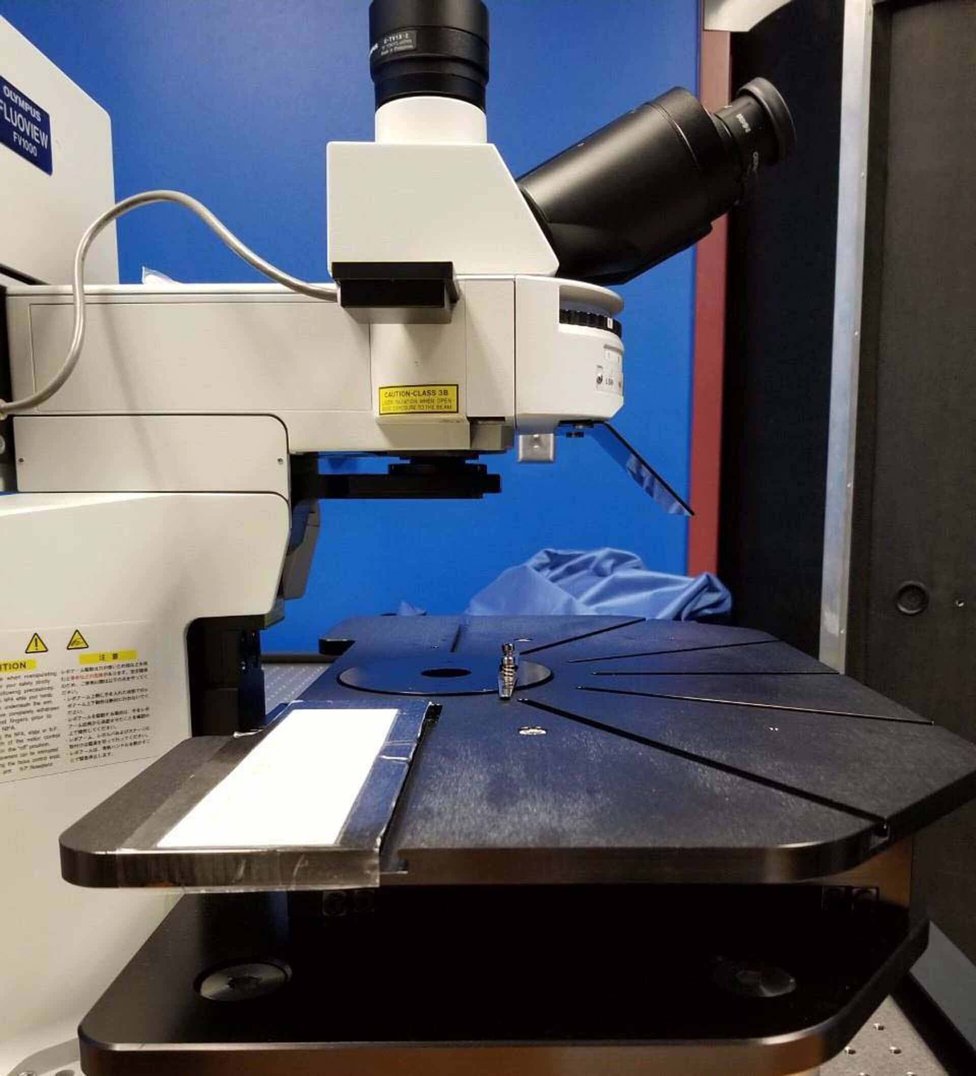

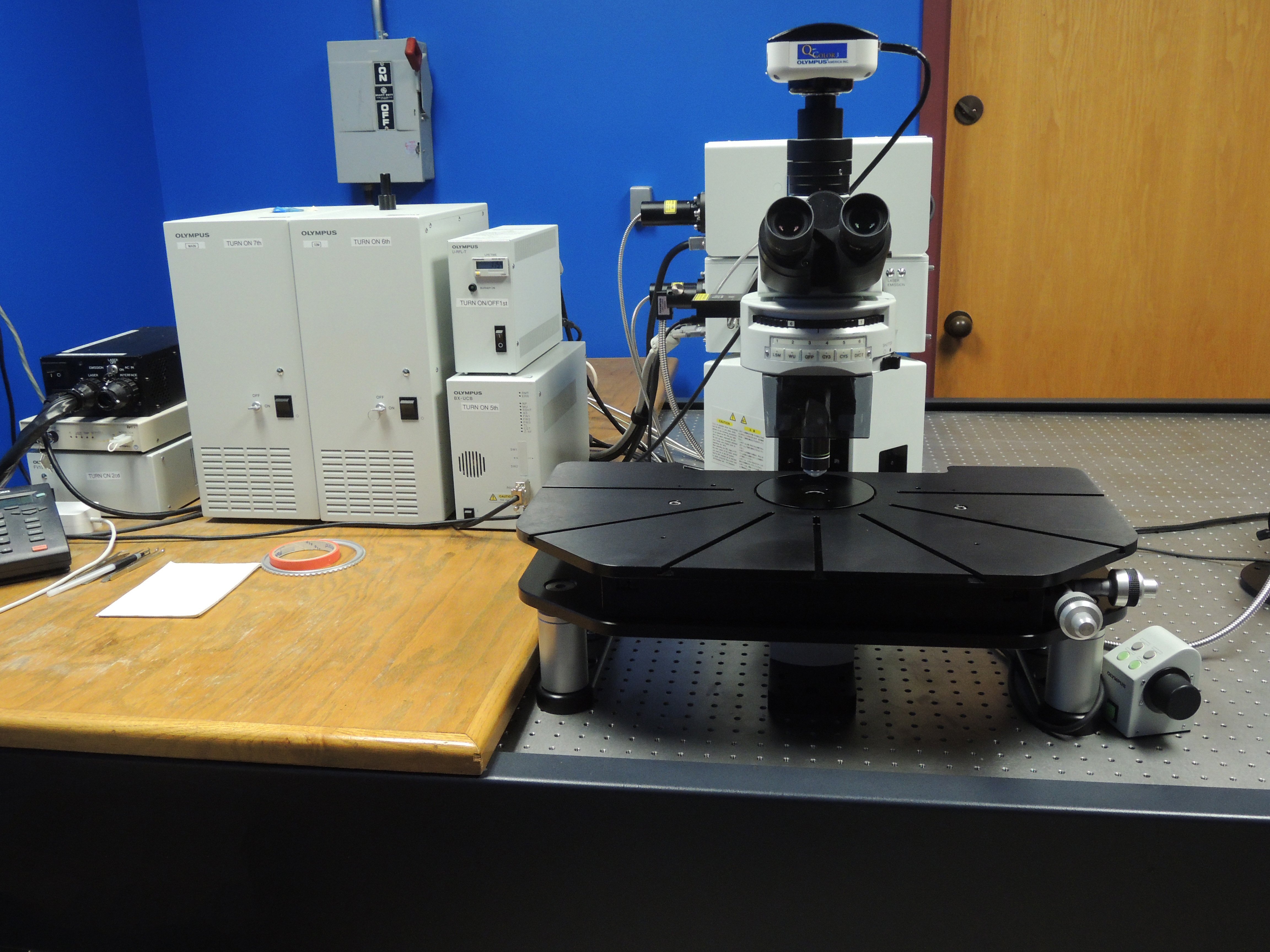


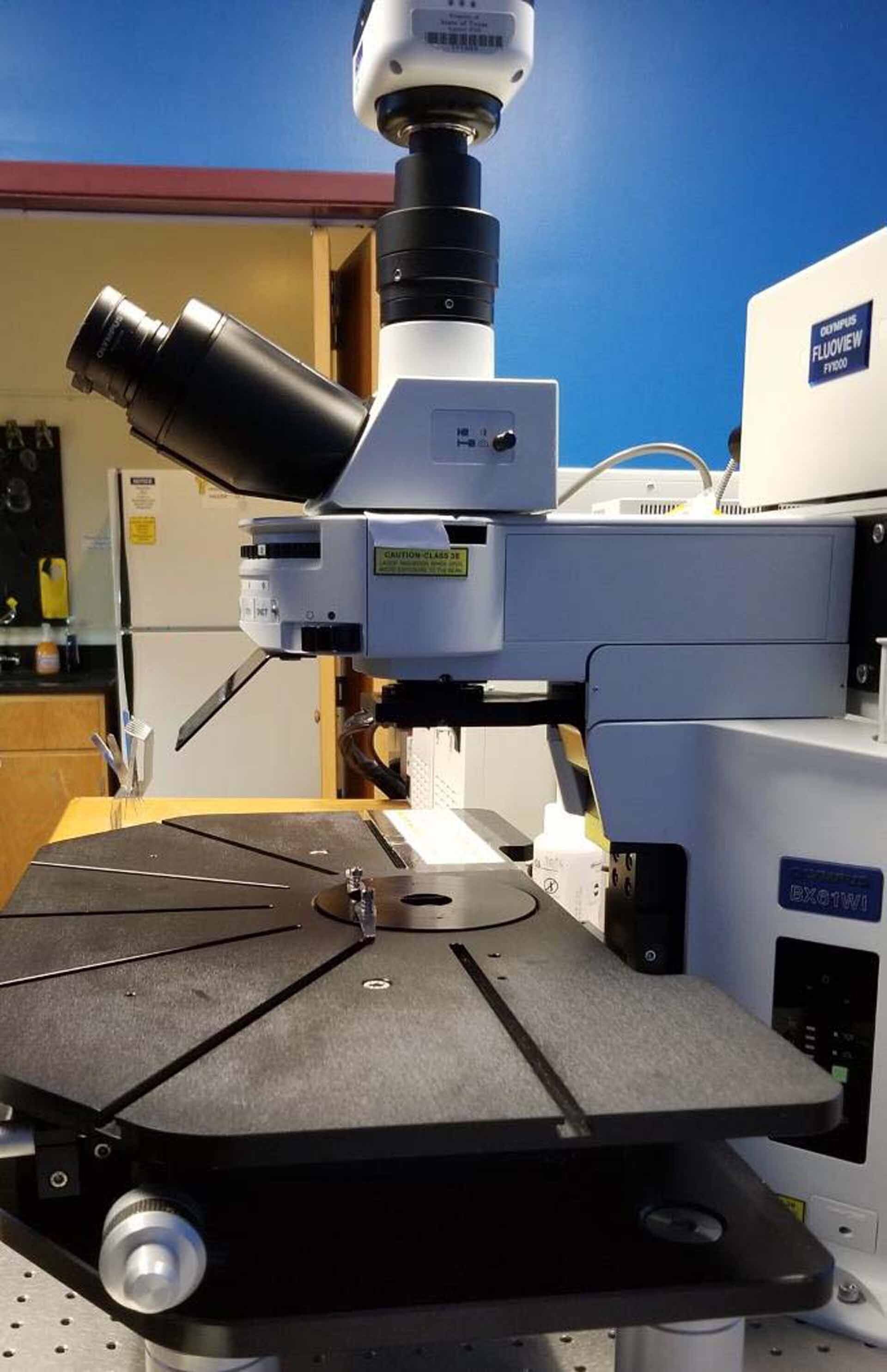














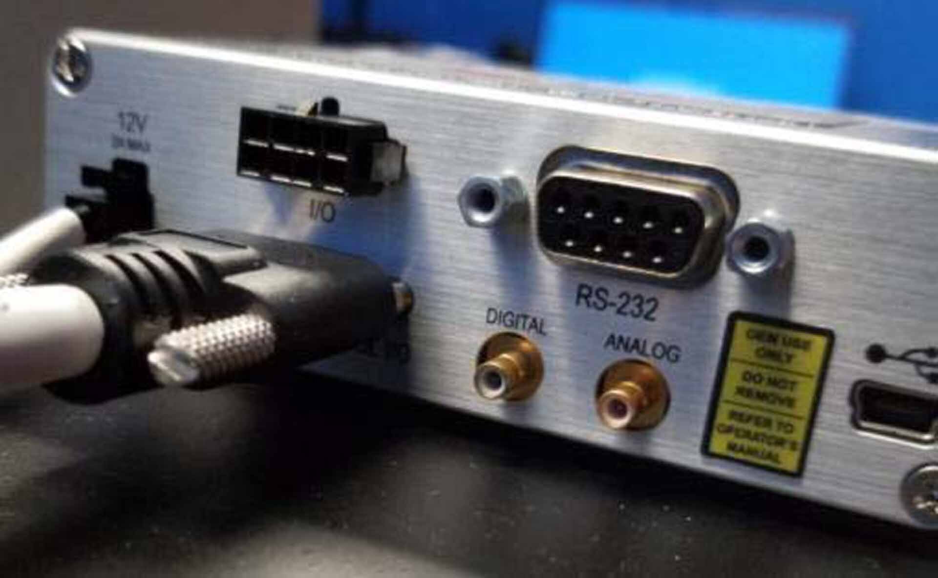


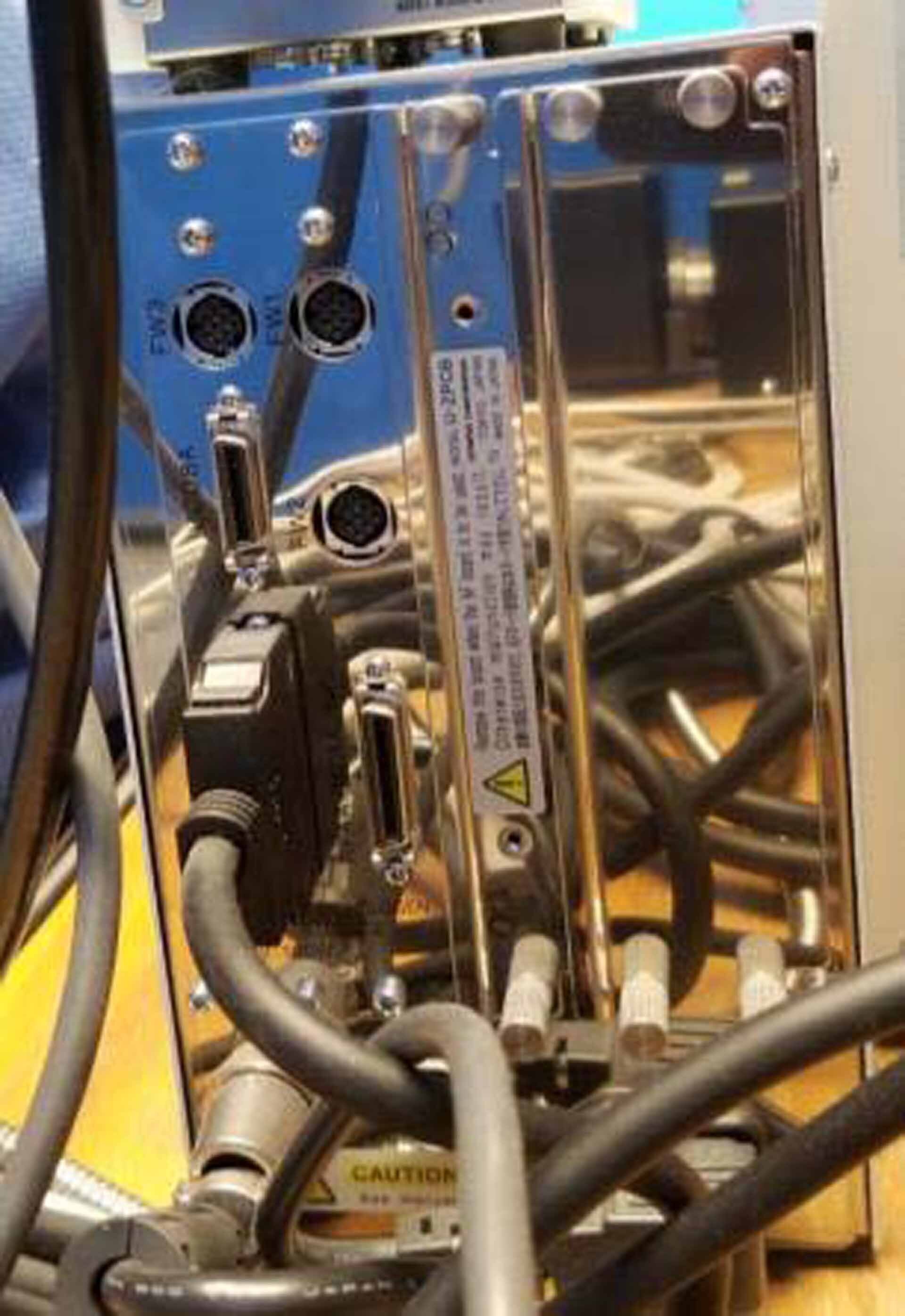








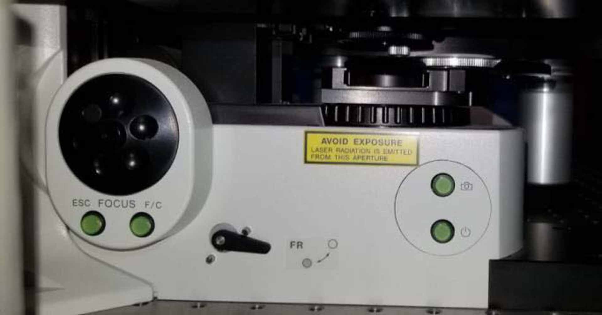

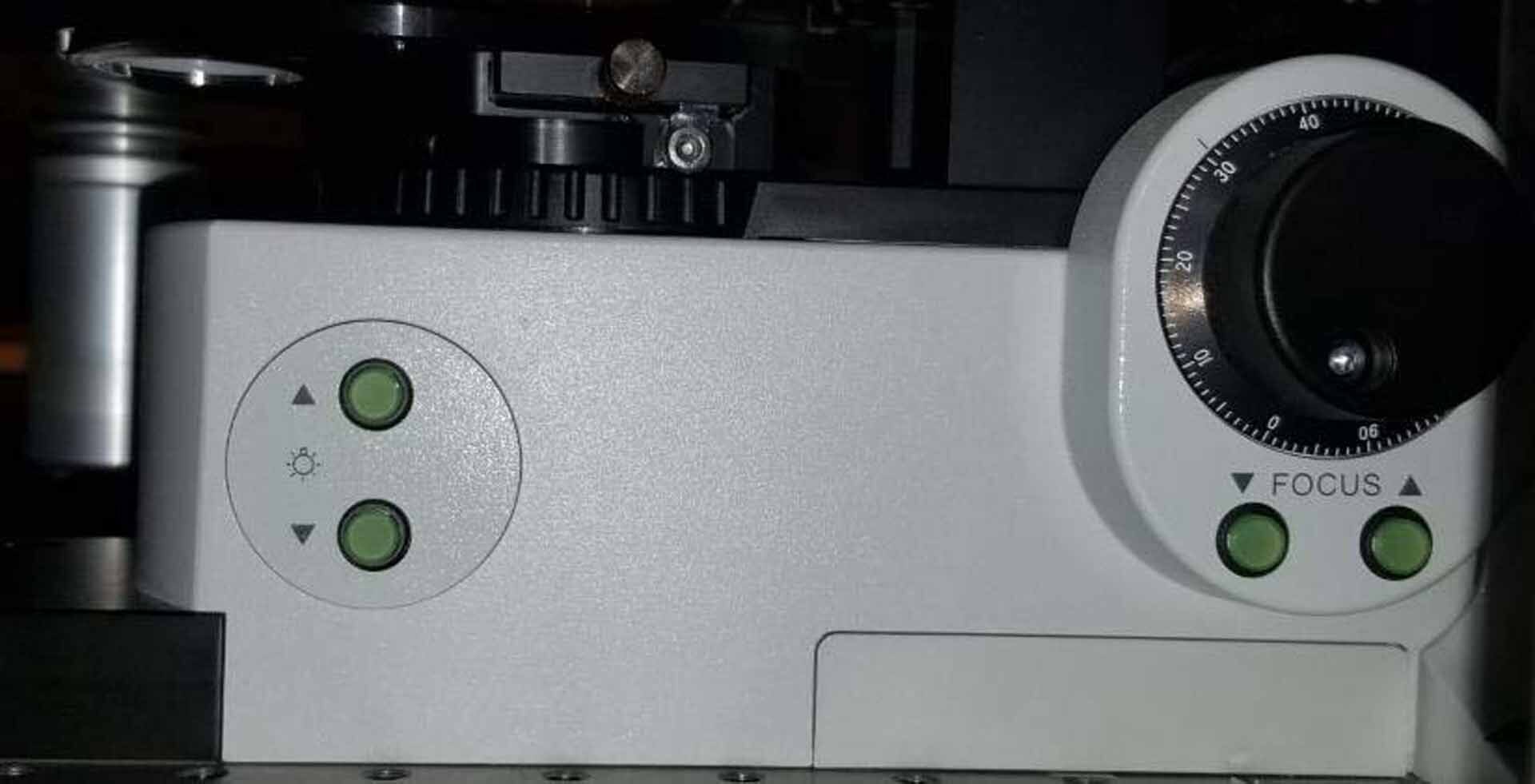


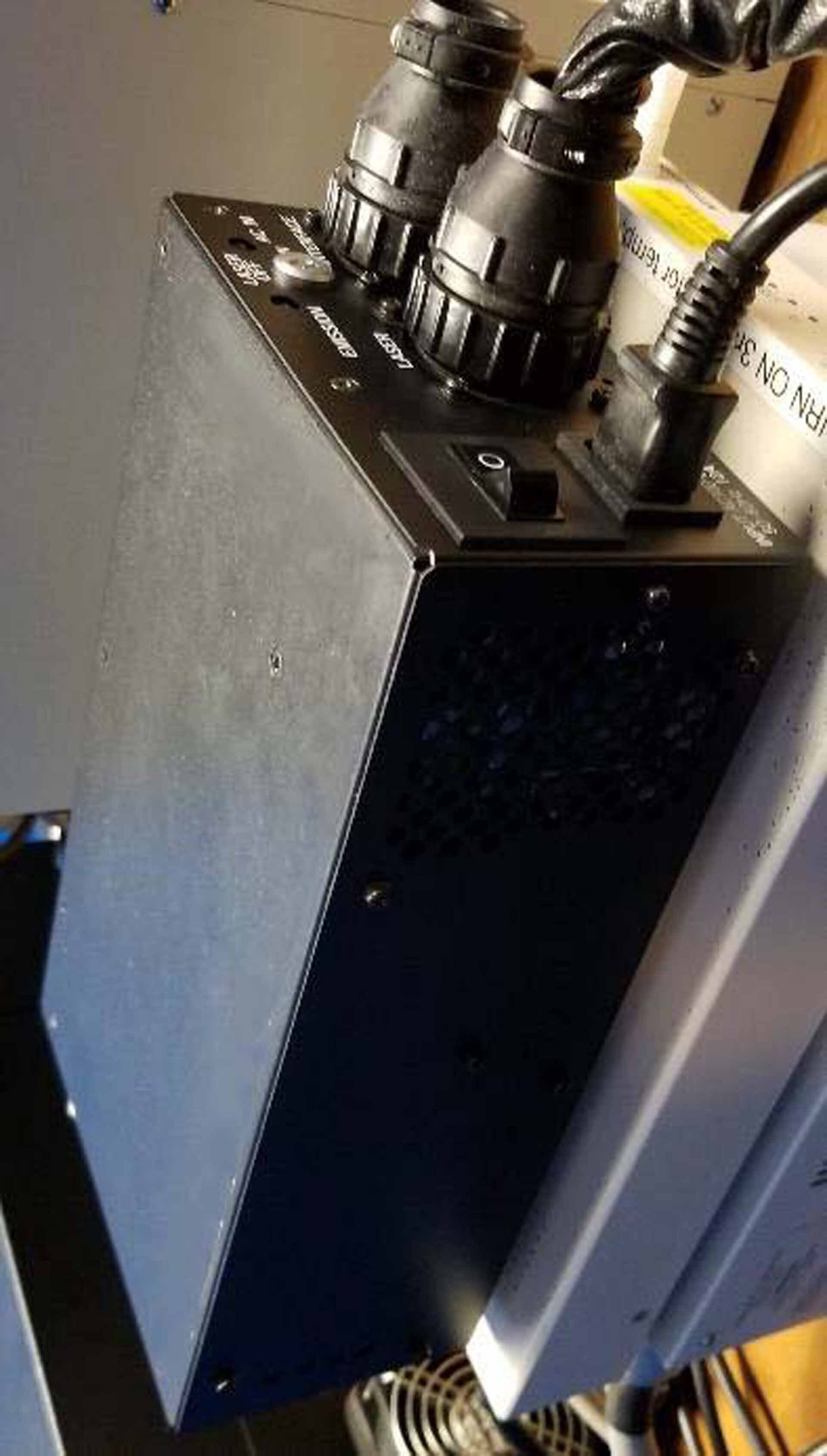






ID: 9305315
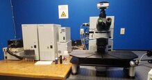
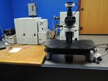
Confocal laser scanning microscope
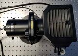
With FV10 TD Transmitted light detector for bright field / DIC Imaging
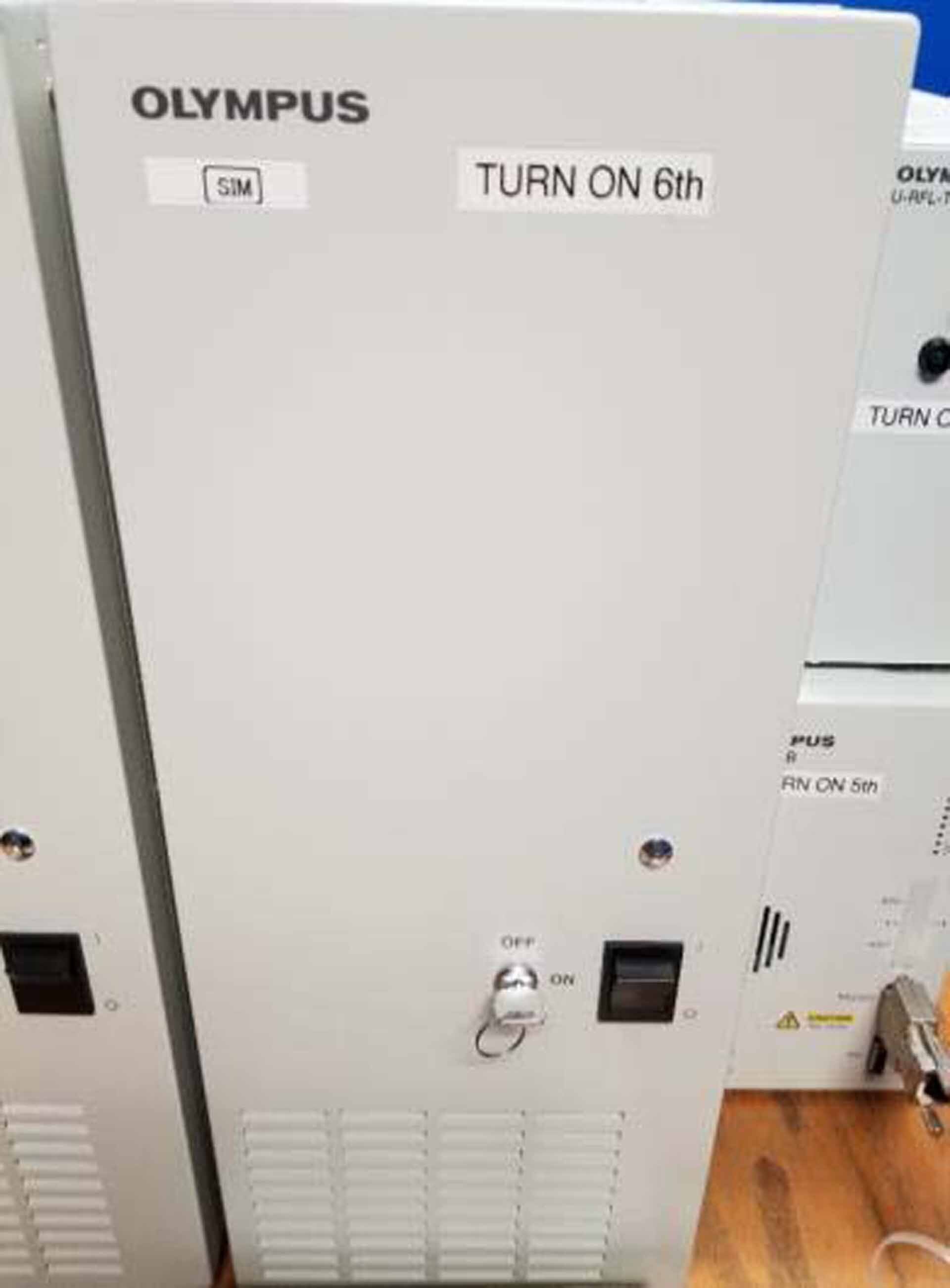
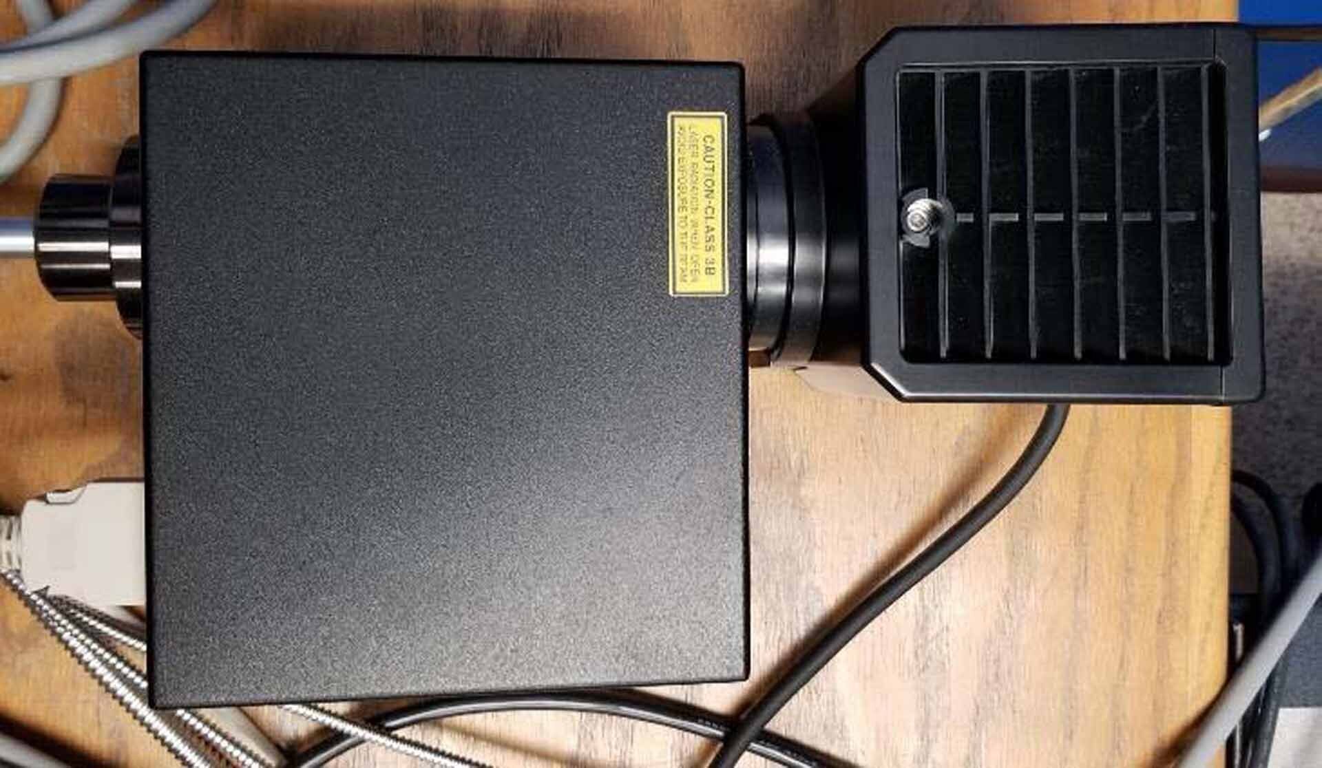
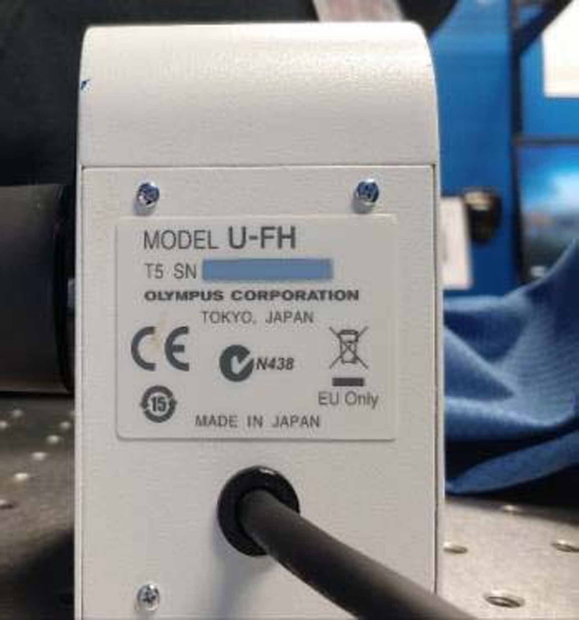
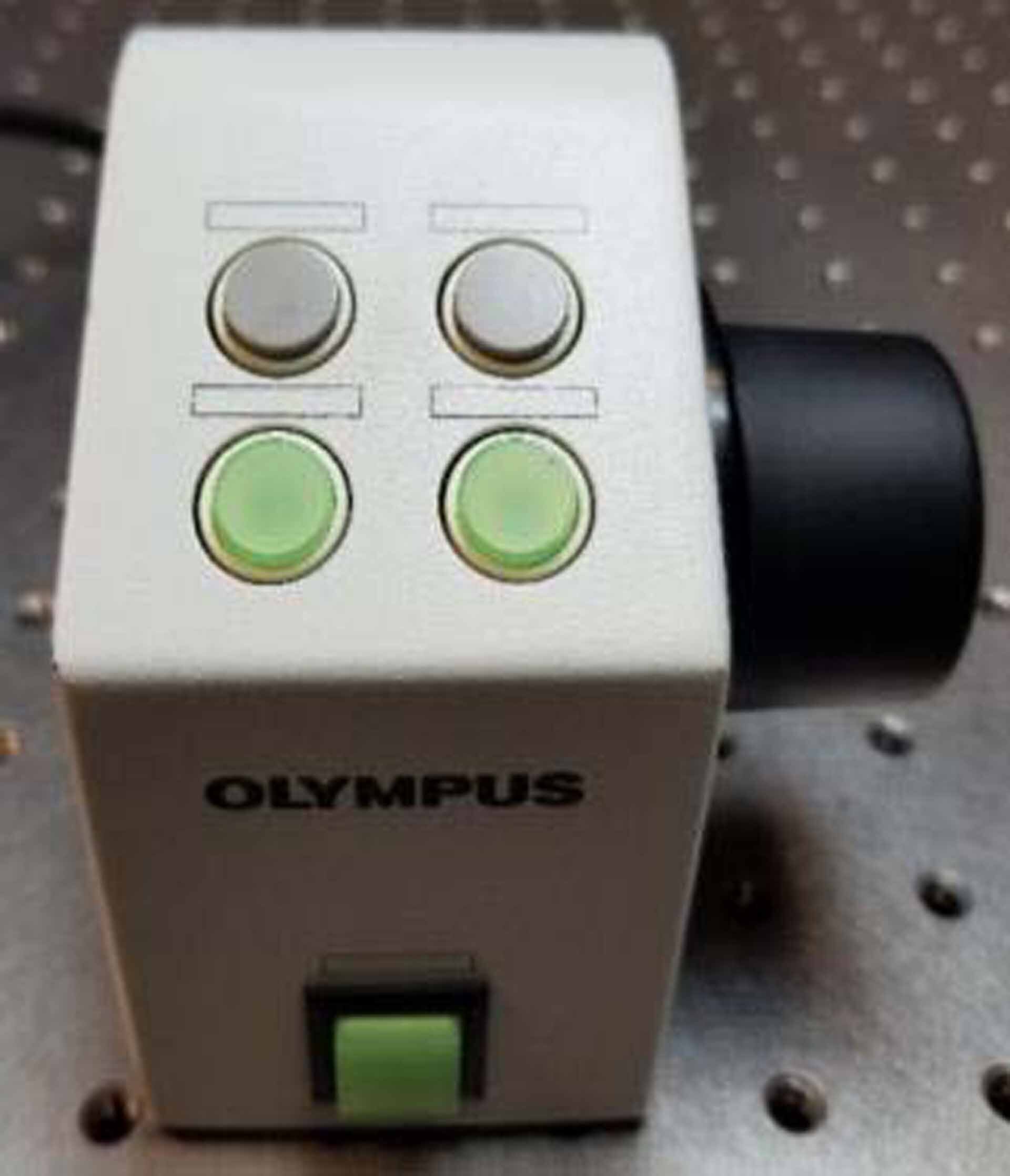
SIM scanner for laser stimulation photo activation / Photo bleaching
F10LMCOMBD2 Laser combiner:
Argon lasers: LD515, LD488, LD458
Diode lasers: LD405, LD559, LD635
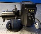
Epi-fluorescence (Mercury lamp)
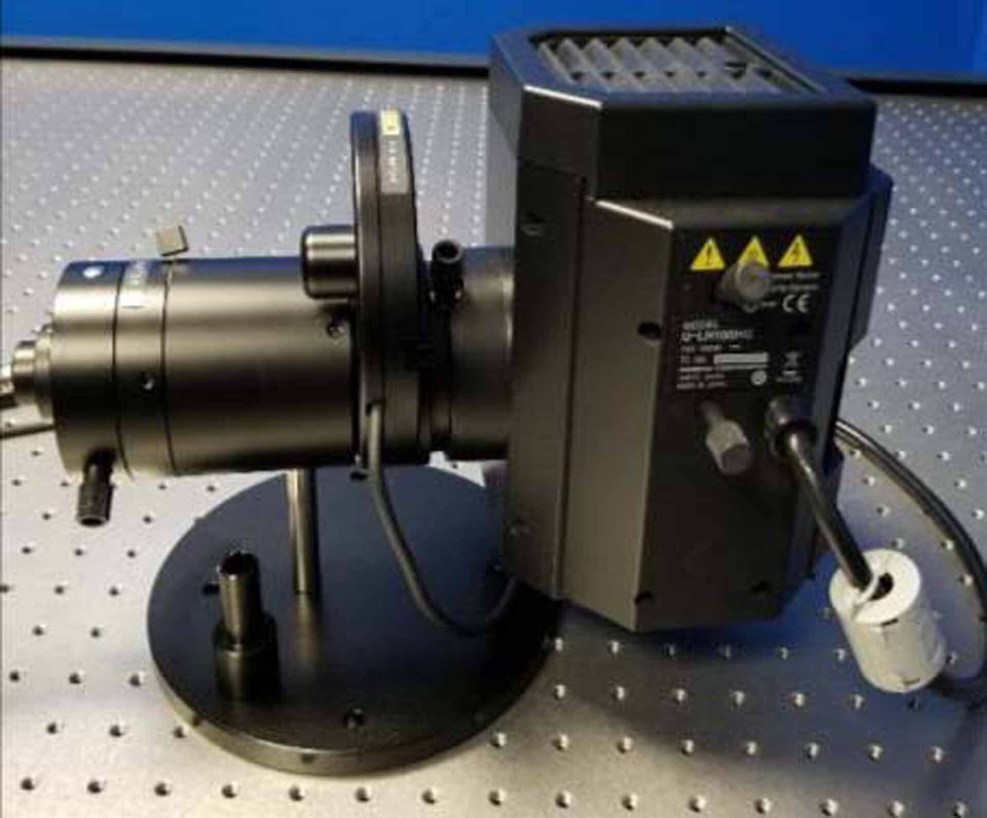
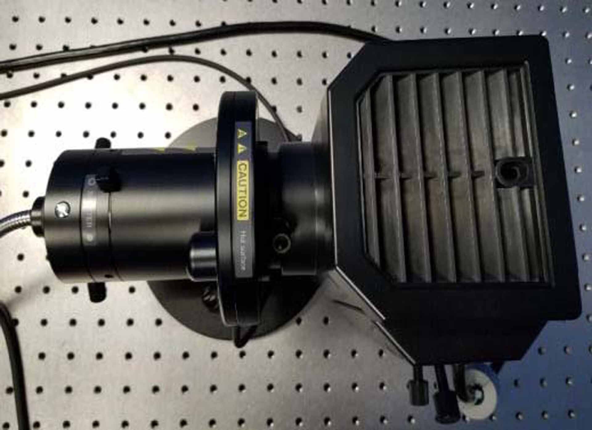
LH100HG Mercury lamp
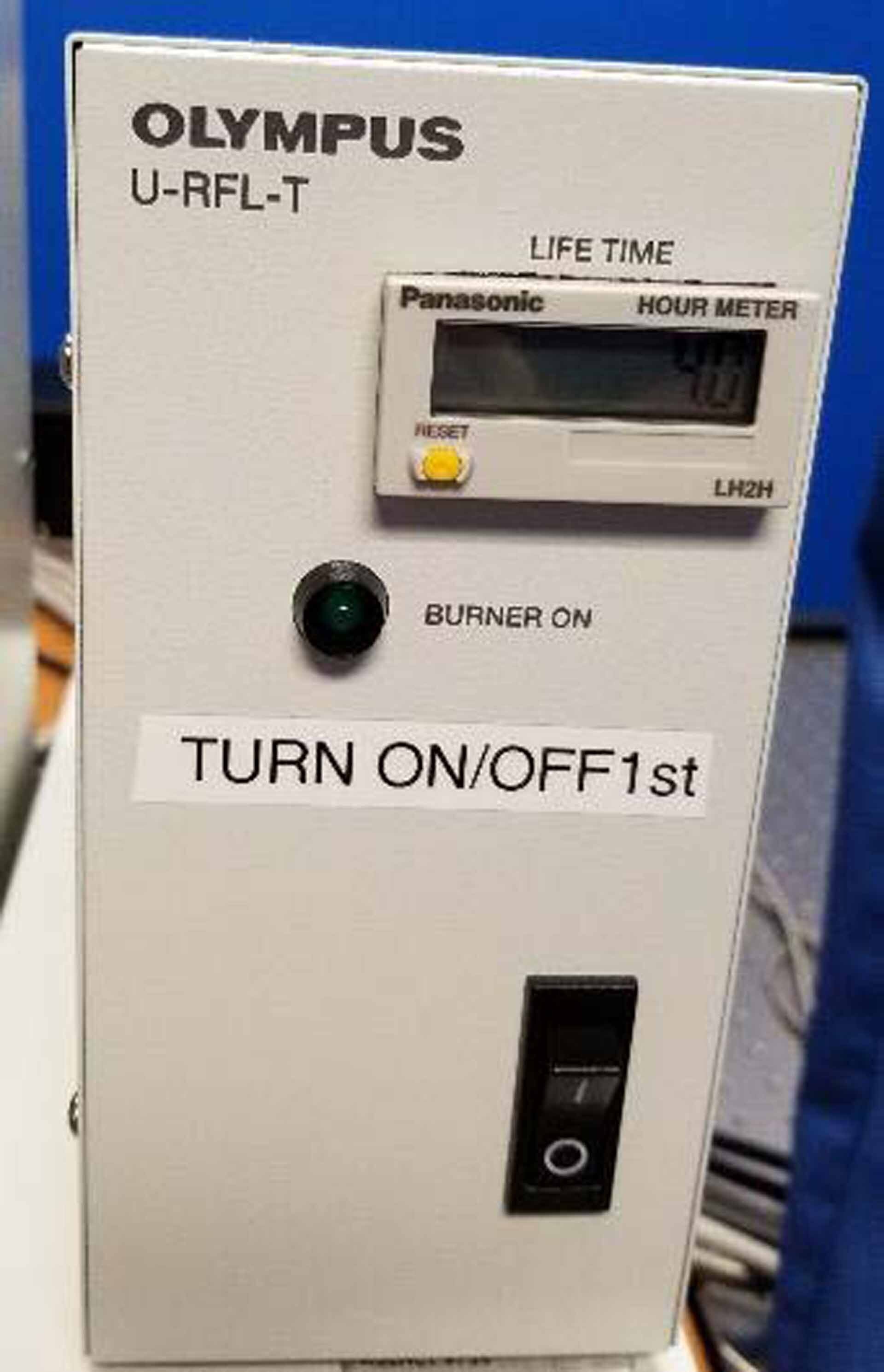
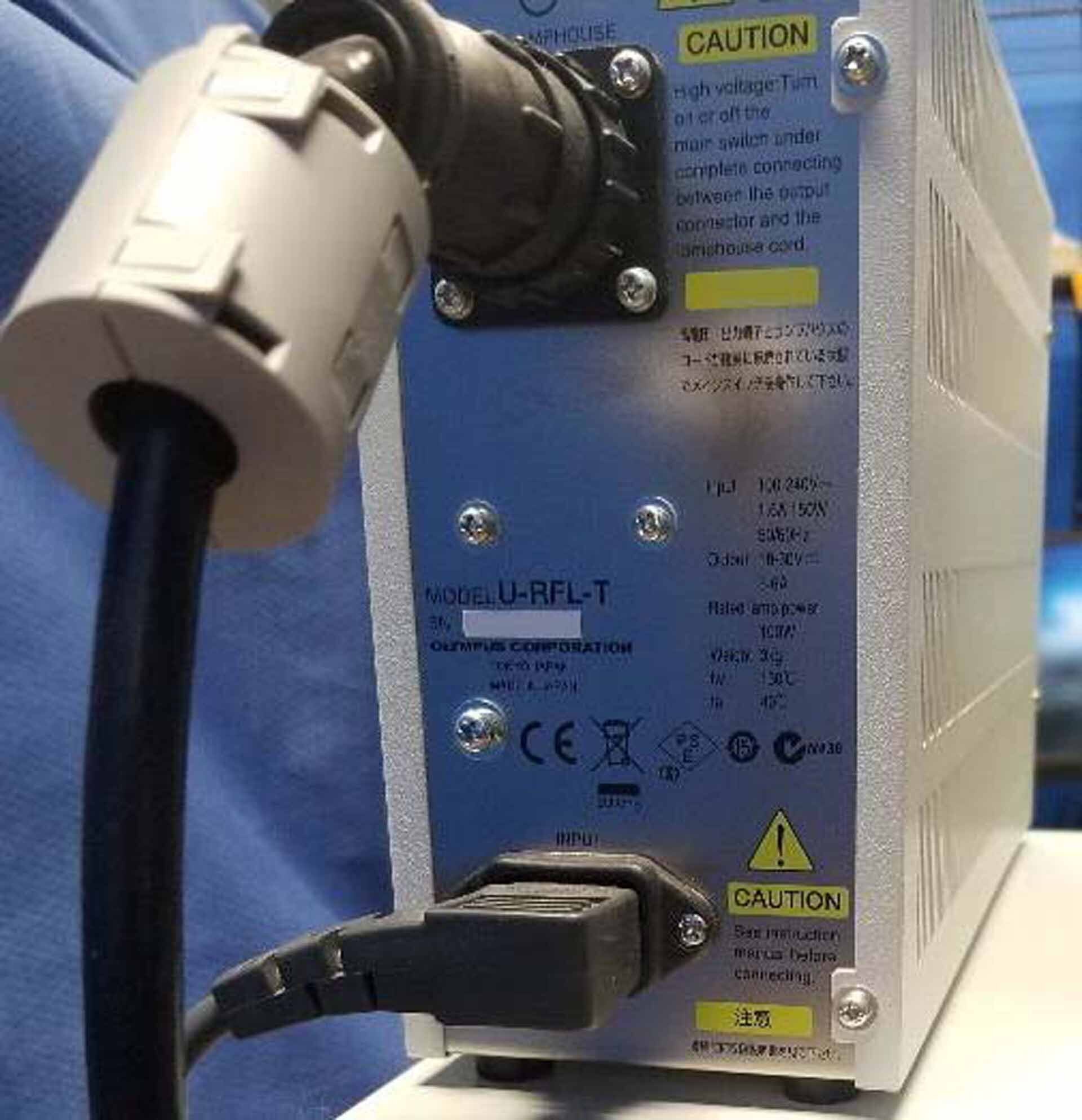
U-RFL-T Power supply for mercury lamp house
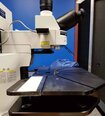
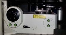
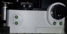
BX61WI Fixed stage with motorized upright microscope
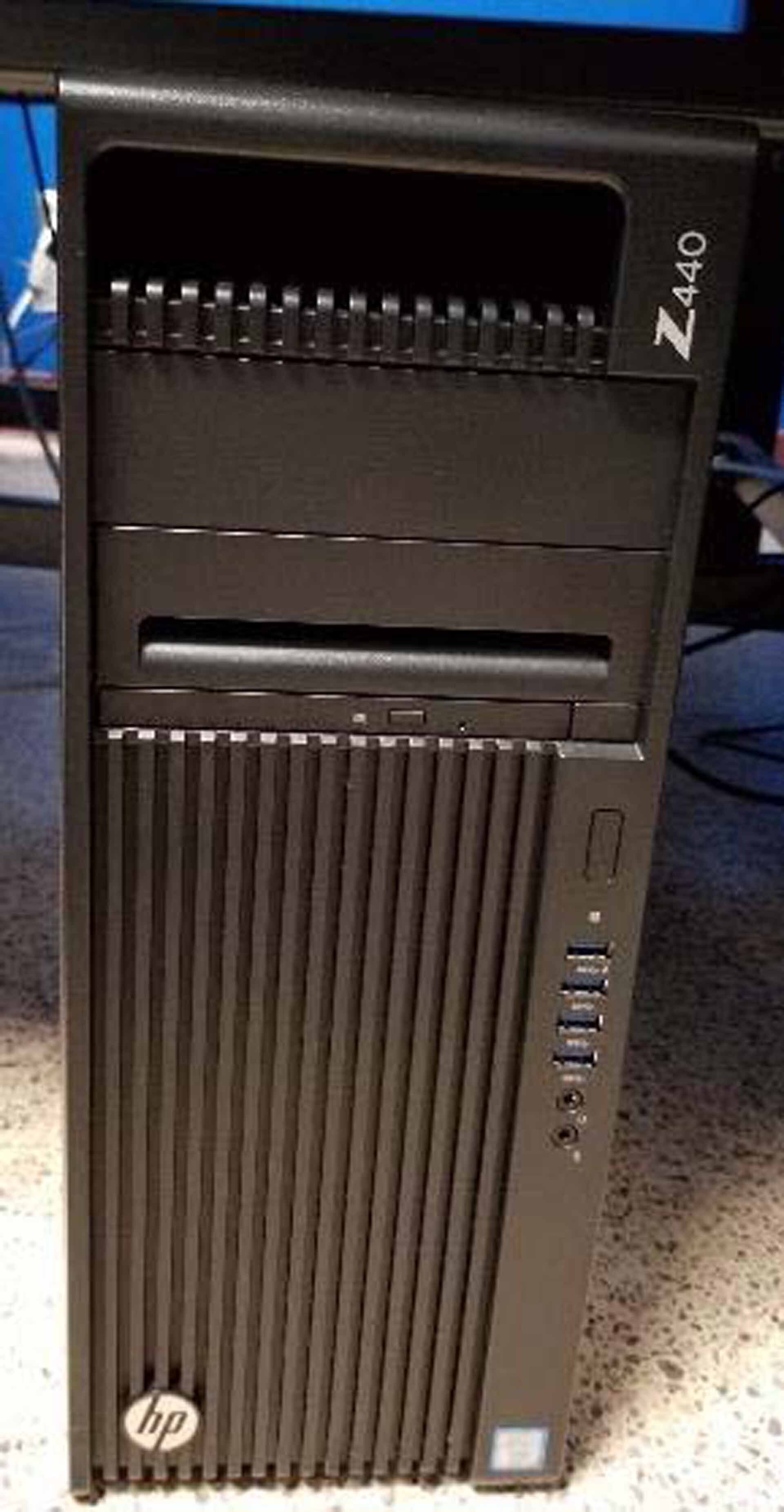
HP Workstation Z440 tower desktop:
INTEL Xeon CPU E5-1650 v3 at 3.50 GHz
RAM: 64.0 GB
System type: 64 Bit
X64 Processor
Keyboard and mouse
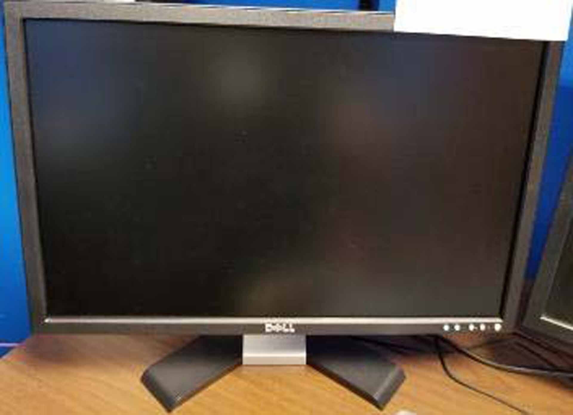
(2) Monitors:
DELL E228WFP
Resolution: 1680 x 1050
Frequency: 60 Hz
DELL P2210
Resolution: 1680 x 1050
Frequency: 60 Hz
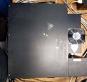
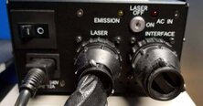
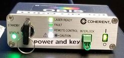
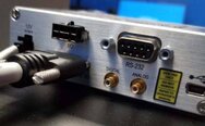
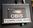
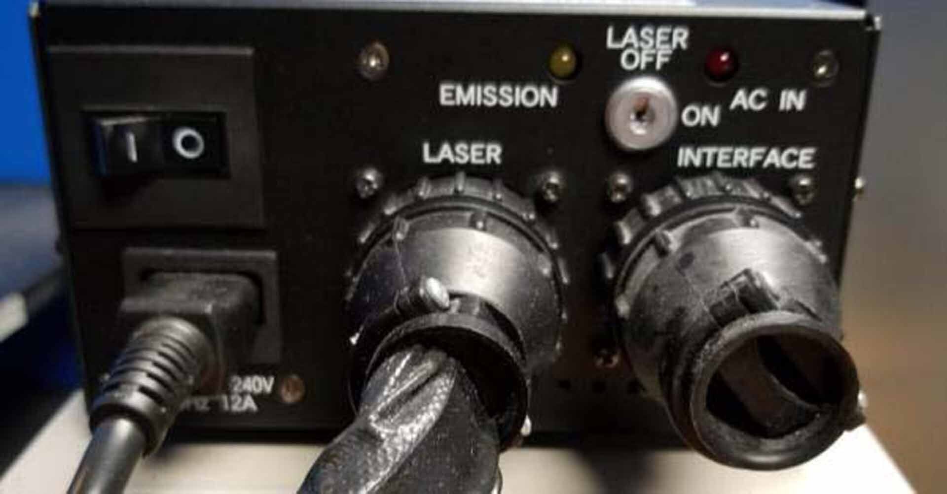
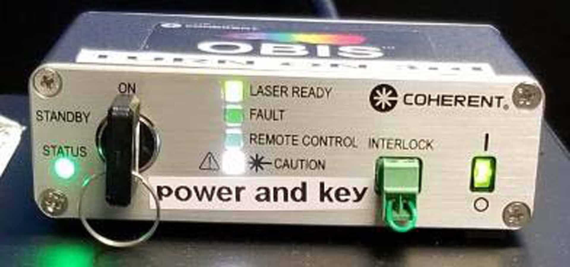
Laser power boxes:
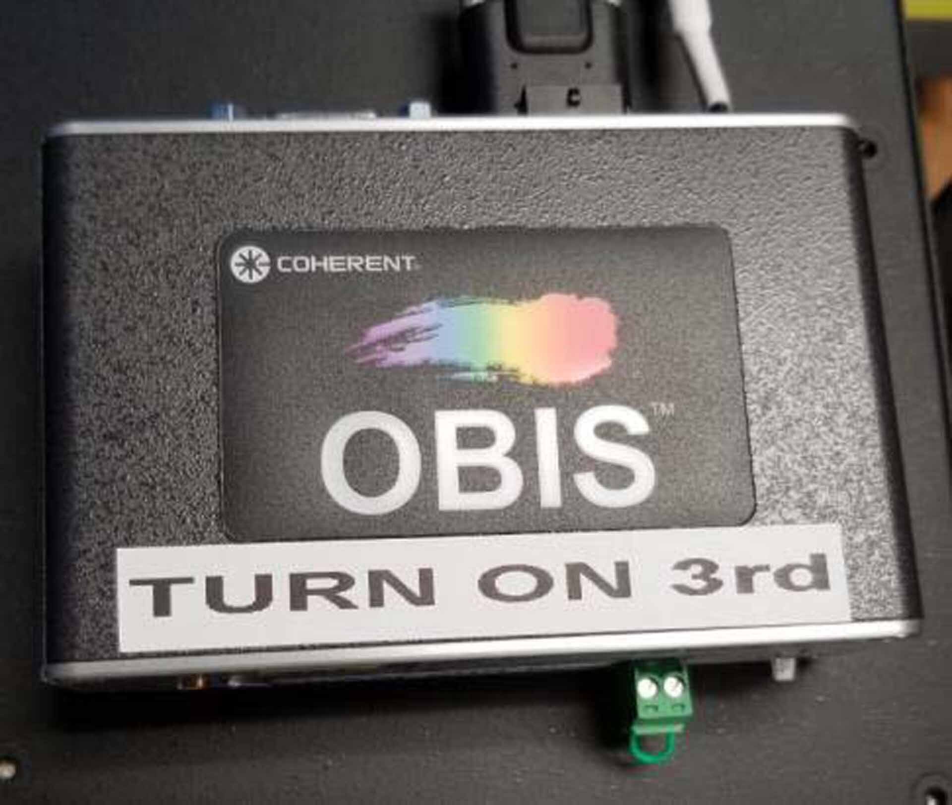
GLS3135
OBIS 561-20 LS CW, 559.8 nm
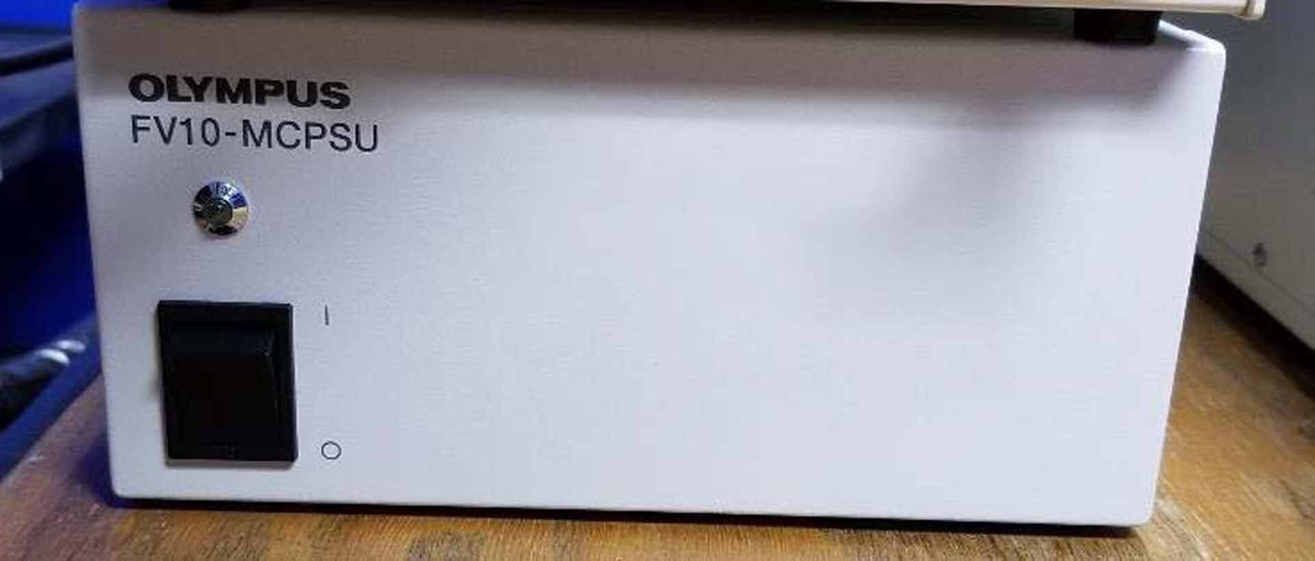
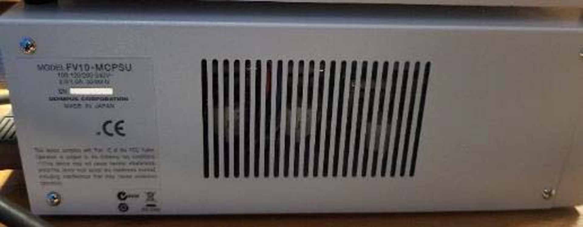
Power supply for FV1000 scan unit
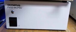
FV10 MCPSU Laser combiner
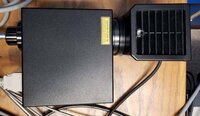
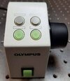
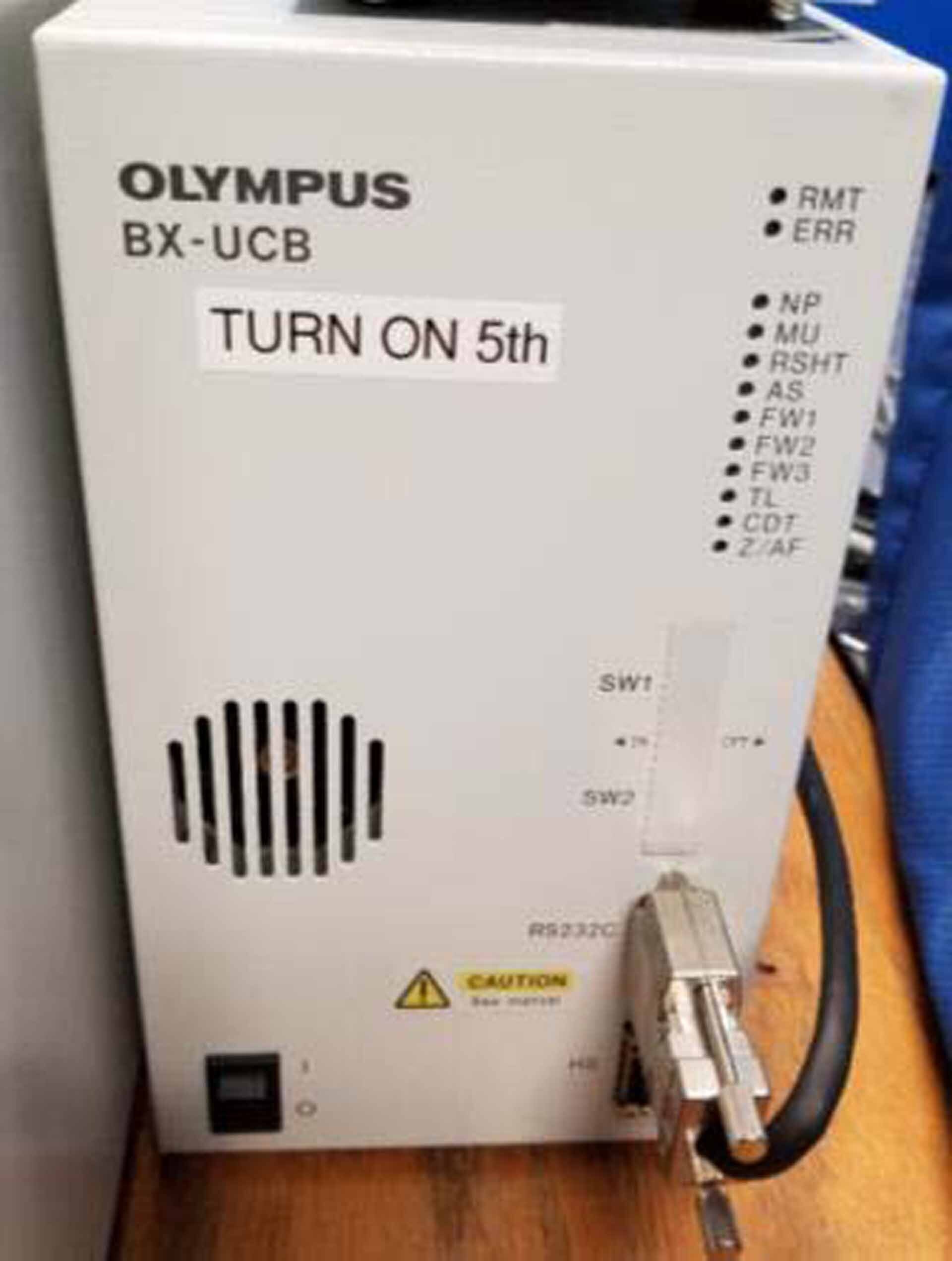
Microscope control box (BX UCB)
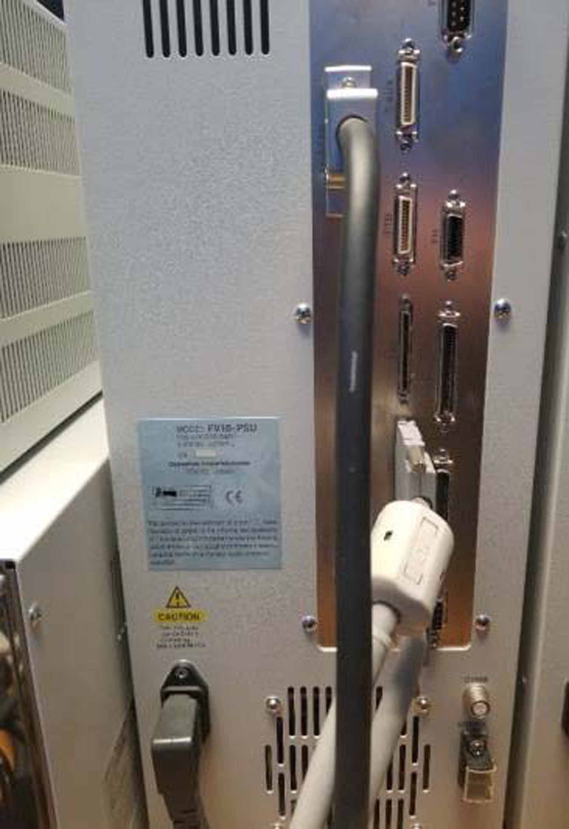
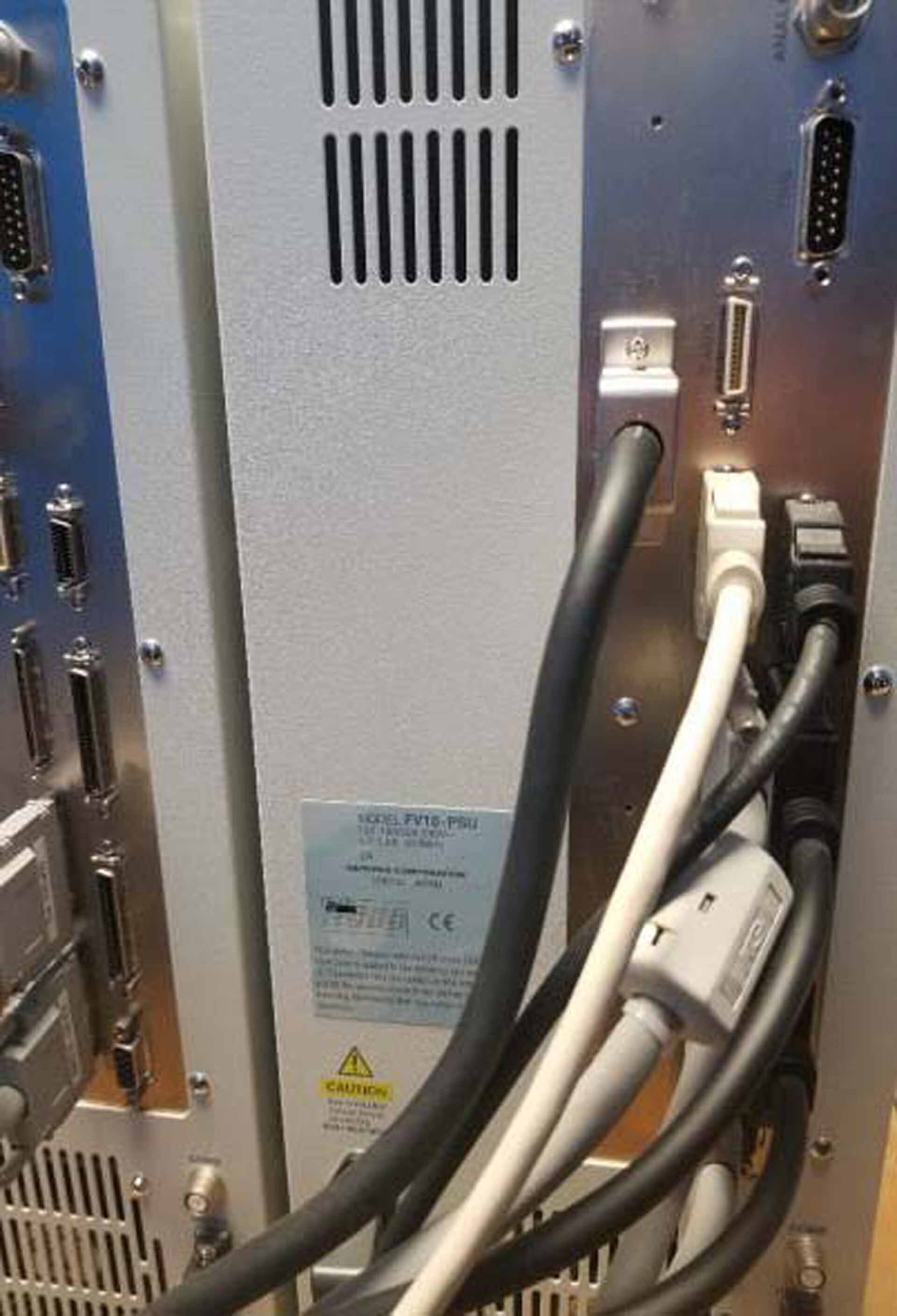
SIM Scanning control box (FV10 PSU)
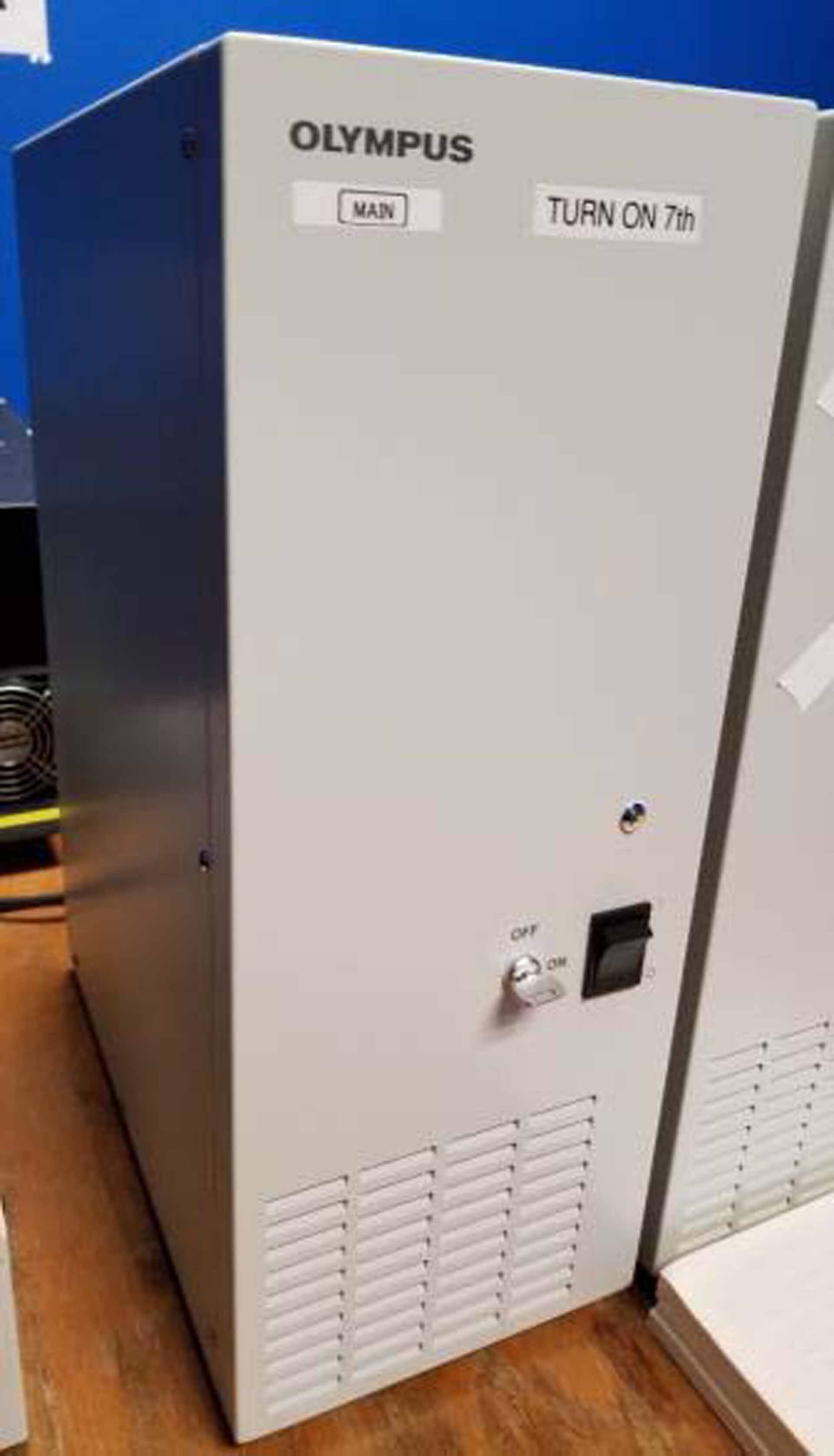
Main scanning control box (FV10 PSU).
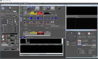
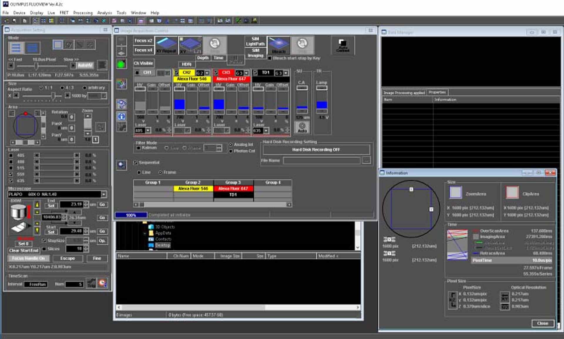
OLYMPUS Fluoview 1000 is a laser scanning confocal microscope that is used in many applications in the life sciences. It has the capability to capture clear, high-resolution images of live cells, as well as provide precise spatial or temporal 3-D imaging. This microscope is powered by a combination of several different light sources, including a laser, mercury arc lamp, and laser diode. The versatile Laser Scanning Unit (LSU) offers several different combinations of illumination, allowing researchers to capture two- and three-dimensional imaging. The specialized light sources work together with sophisticated optical systems to generate powerful fluorescence, which is used to collect and analyze images. The microscope is equipped with a powerful software suite featuring advanced image processing that can be used to quantify or map the data. This confocal imaging system can also be integrated with other optical components, such as a widefield microscope or multiphoton microscope. The ultra-compact, ergonomic design of Fluoview 1000 makes it a great choice for any laboratory. It features a large working distance, allowing for much more flexibility when working with the specimen. The sample can be easily moved, rotated, and zoomed in the field of view. The microscope also offers an extensive collection of filters and excitation sources, allowing the user to explore a range of fluorescence microscopy techniques. Additionally, OLYMPUS Fluoview 1000 can be used to study a wide variety of samples, from normal tissue sections to complex, thick specimens. To ensure precise image acquisition, Fluoview 1000 can be tailored to meet the needs of a large variety of applications. The advanced imaging system also offers intuitive control of the microscope, as well as a user-friendly interface with tools for image analysis. It is ideal for studying a wide range of topics, from virus life cycles to cell signaling and beyond.
There are no reviews yet
