Used FEI / PHILIPS Microscope #293816314 for sale
URL successfully copied!
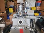
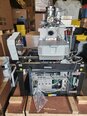
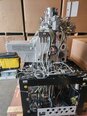
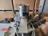


Tap to zoom

ID: 293816314
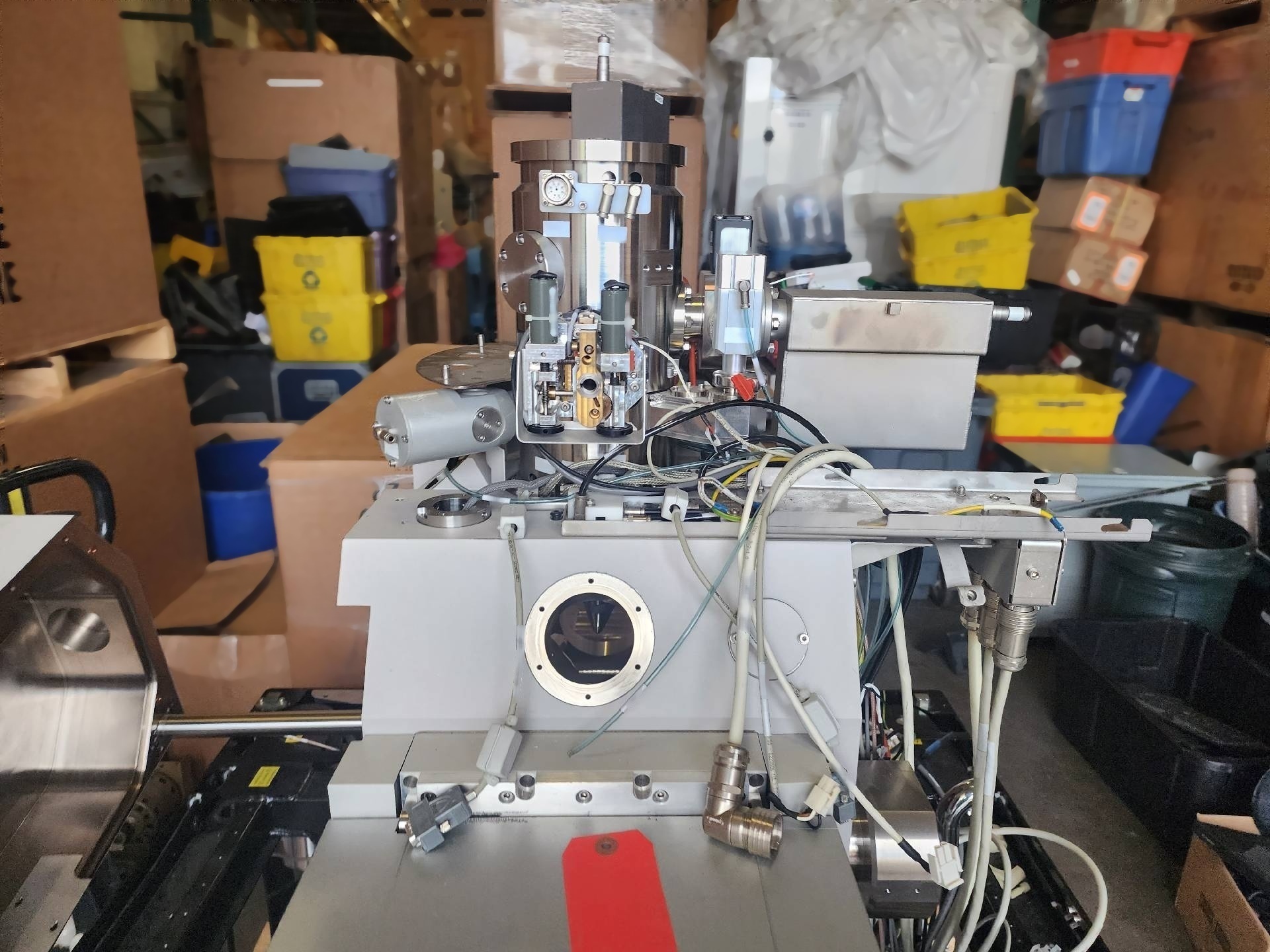
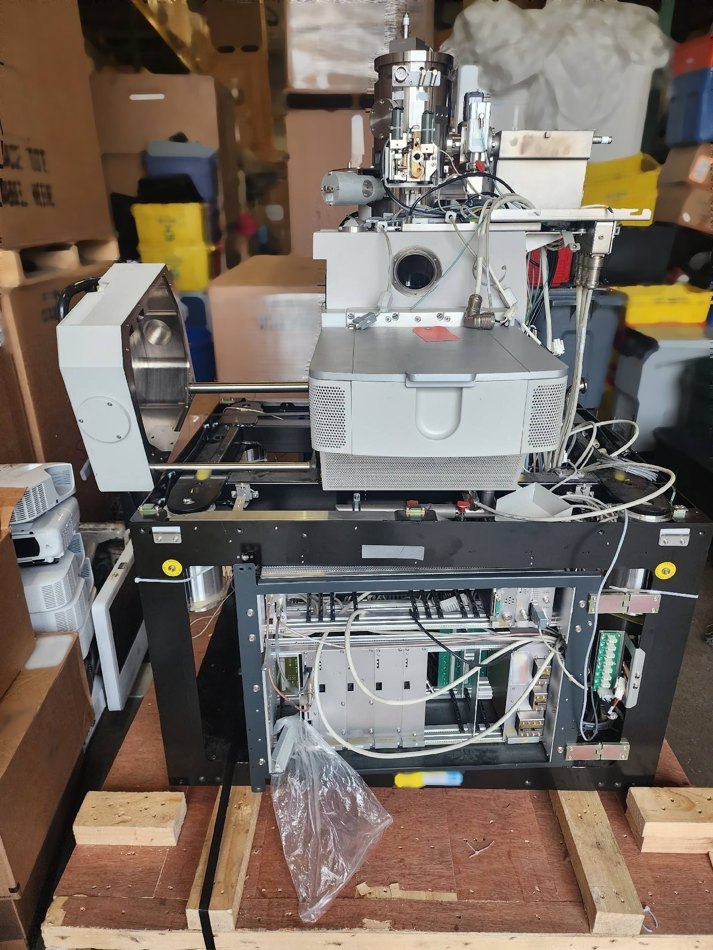
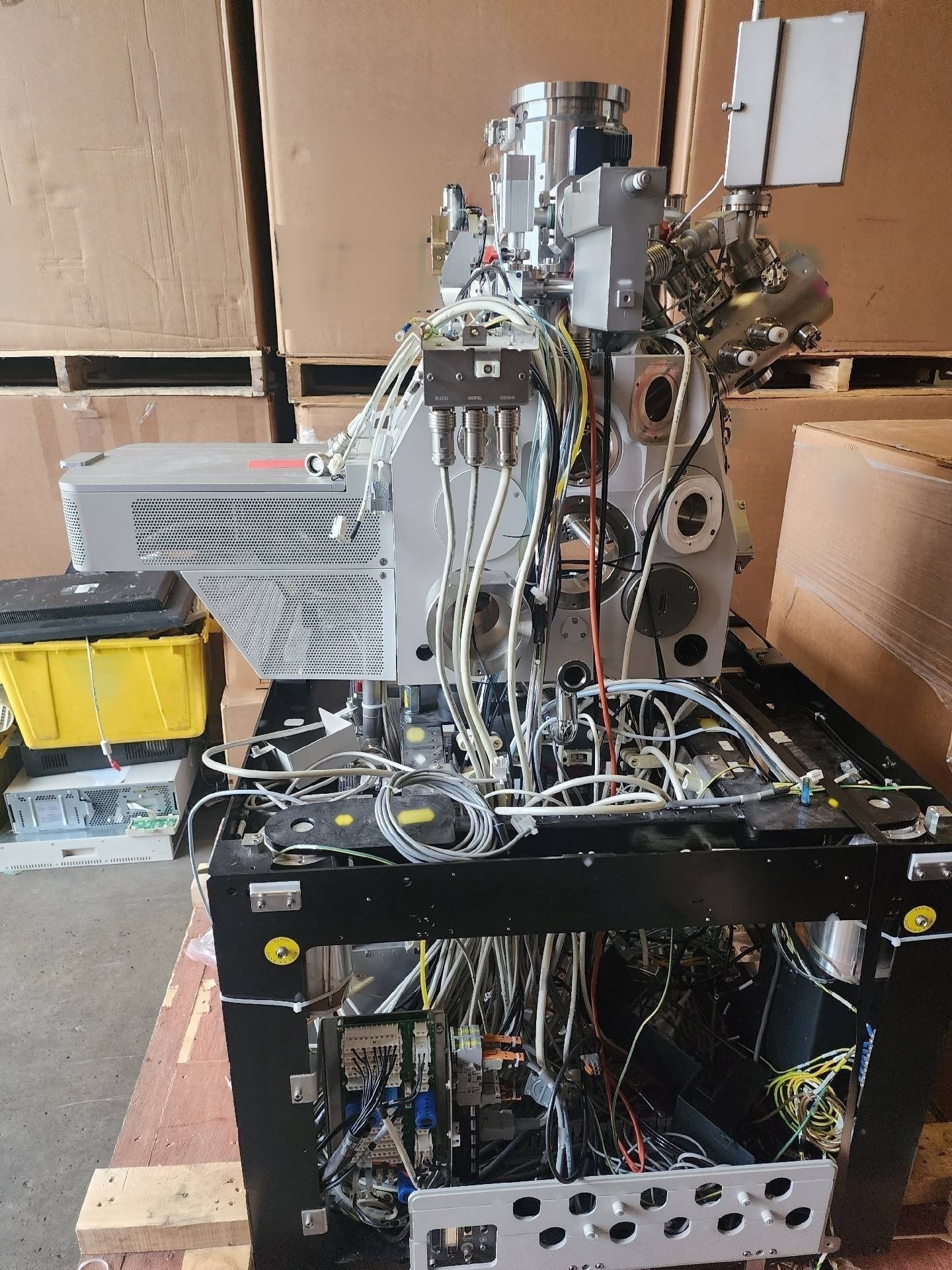
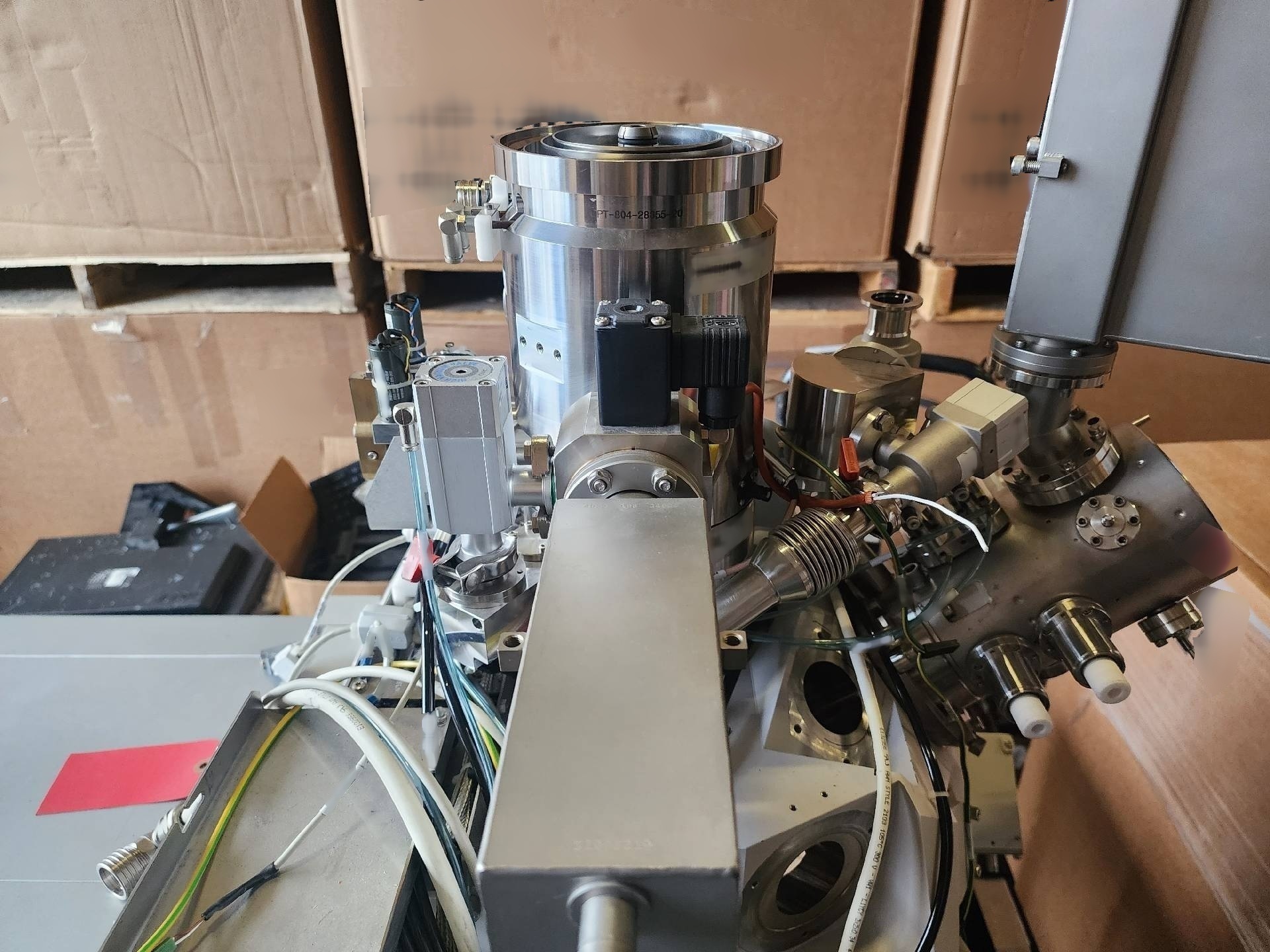
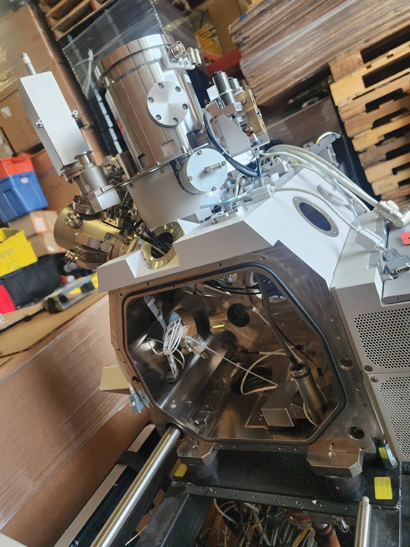
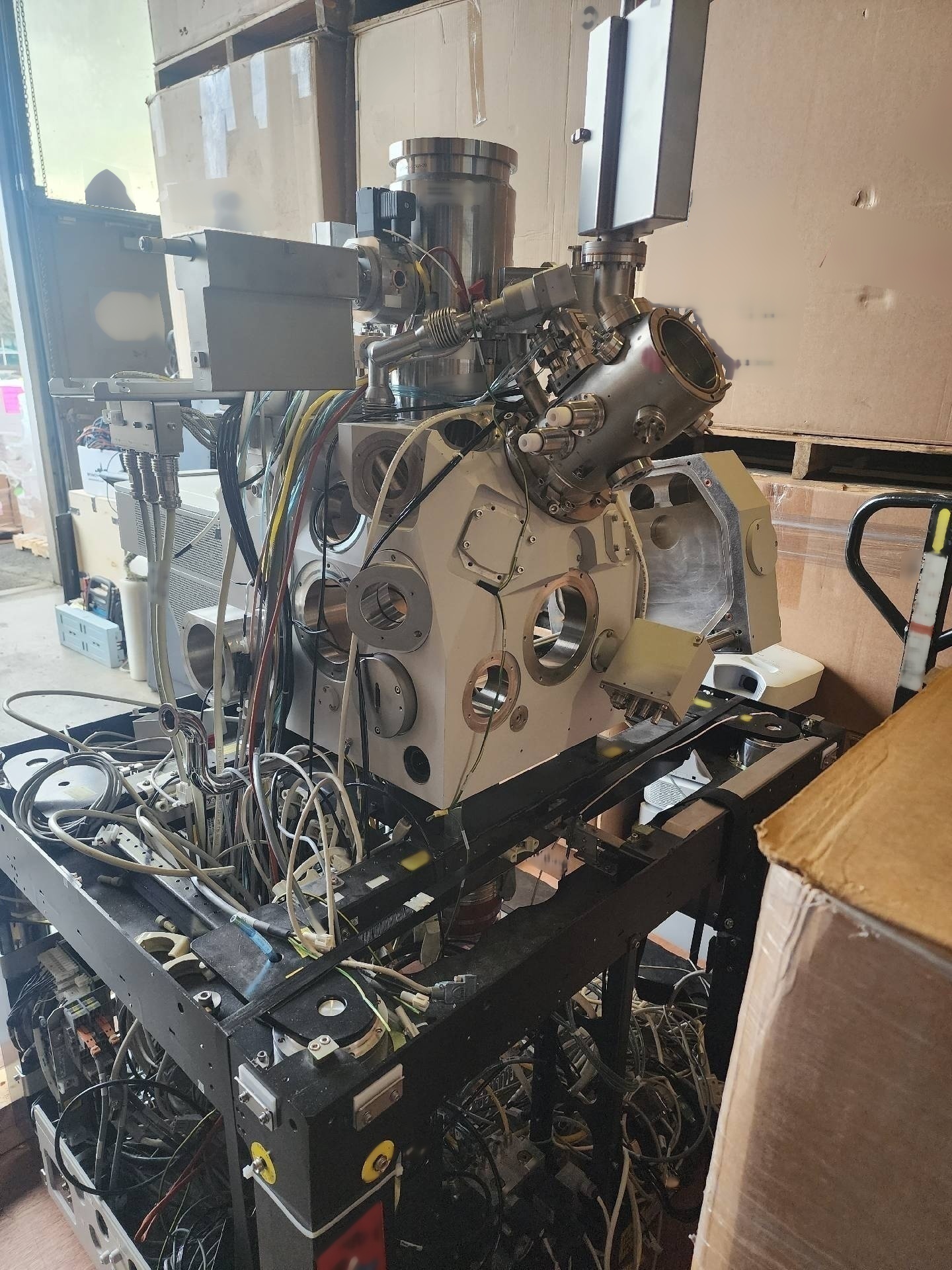
FEI scanning electron FEI / PHILIPS Microscope (SEM) is an advanced imaging instrument that enables researchers to visualize the microstructure of samples with extremely high resolution and magnification. This powerful analytical tool utilizes a focused beam of electrons to create detailed images of sample surfaces, providing valuable insights into their topography, composition, and chemical properties. At the heart of PHILIPS SEM is an electron gun, which generates a beam of high-energy electrons that is focused and accelerated towards the sample surface. The electron beam interacts with the sample, resulting in various signals that can be detected and used to create an image. One of the key advantages of SEM imaging is its ability to achieve much higher resolution compared to conventional optical microscopes, making it an indispensable tool for a wide range of scientific disciplines. FEI / PHILIPS SEM is equipped with a variety of detectors that can capture different types of signals generated by the interaction of electrons with the sample. The secondary electron detector, for example, is commonly used to produce images of the sample's surface topography, providing detailed information about its features and textures at the nanoscale level. In addition, the backscattered electron detector can be utilized to generate images that are sensitive to variations in the sample's composition, offering insights into its chemical composition and elemental distribution. Aside from imaging capabilities, FEI SEM also features advanced analytical tools that enable users to perform a wide range of characterization techniques. Energy-dispersive X-ray spectroscopy (EDS) is a commonly used technique that allows researchers to analyze the elemental composition of samples by detecting the characteristic X-rays emitted when the sample is bombarded with electrons. This provides invaluable information about the distribution and concentration of elements within the sample, aiding in materials analysis and identification. Another powerful analytical tool available in PHILIPS SEM is electron backscatter diffraction (EBSD), which enables researchers to study the crystallographic structure of samples at the microscopic level. By analyzing the patterns formed by electrons diffracted from crystalline regions within the sample, EBSD can provide information about grain orientation, phase identification, and grain boundaries, facilitating advanced materials research and development. FEI / PHILIPS SEM is designed to be user-friendly and versatile, allowing researchers to customize imaging and analytical parameters to suit their specific research needs. Its intuitive software interface enables easy control of imaging settings, data acquisition, and image processing, making it an ideal tool for both novice users and experienced researchers alike. In conclusion, FEI scanning electron FEI Microscope is a cutting-edge instrument that offers unparalleled imaging and analytical capabilities for studying the microstructure of materials at the nanoscale level. With its high resolution, advanced detectors, and versatile analytical tools, PHILIPS SEM is a valuable asset for a wide range of scientific and technological applications, including materials science, geology, biology, and nanotechnology.
There are no reviews yet