Used HITACHI S-3000N #9208239 for sale
URL successfully copied!
Tap to zoom


ID: 9208239
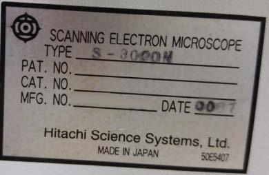
Vintage: 2000
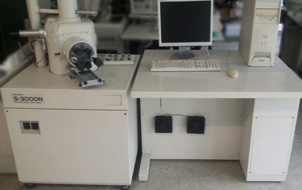
Scanning Electron Microscope (SEM)
Option: W4010 Water chiller
Magnification: 15x to 300000x (143 steps)
Resolution:
Secondary electron image resolution:
3.0 nm (Vacc 25 kV, high vacuum mode)
Back-scattered electron image resolution:
4.5 nm (Vacc 25 kV, variable pressure mode)
Voltage: 0.3 - 30 kV
Specimen size: 150 mm Diameter maximum
View monitor: 17" LCD Type
Operation: Mouse and sub control pad
PC: PC/AT Compatible
Operating system: Windows NT
Electron gun:
W (Tungsten)
Hairpin type
Stages:
X-Axis: 0 - 100 mm
Y-Axis: 0 - 50 mm
Z-Axis: 5 - 40 mm
Tilt: -5 ~ +60°
Rotation: 360° (Non step)
Vacuum pumps:
(2) Rotary pumps
Diffusion pump
(2) HITACHI CuteVac VR16L-K Rotary pumps
Control system:
TV Fast mode
TV Flow mode
1-4 Step slow mode
Reduce mode
Power / Utility:
PCW: 1-1.5 liter/min
Pressure: 0.5-1 kg/cm³
Pressure dry air (PDA): 5 kg/cm³
AC 100 V, Single phase, 5 kVA within GND
2000 vintage.
HITACHI S-3000N is an advanced scanning electron microscope (SEM) designed for use in advanced materials research and analysis. The microscope is equipped with a high resolution flat field optics equipment, allowing for sub-micrometer resolution. It also employs an in-column energy filter allowing for fine tuning of imaging parameters. The device also has a high voltage power supply with an accuracy of 0.01 volts, allowing for precise and accurate imaging. HITACHI S-3000 N is capable of performing both standard and variable-pressure sample viewing. High sensitivity secondary electron imaging and backscatter electrons allows for the observation of various features on sample surfaces. Additionally, the device is synchronized with other imaging systems, making it possible to conduct correlative research. The device is outfitted with an X-ray analyzer and the results can be analyzed via an automated optimization algorithm. This allows for the highest quality SEM imaging and sample analysis. Additionally, the microscope includes an automatic stage control system and an automated edge detection feature, allowing for consistent and repeatable imaging. The unit includes an automated parameter optimization process, allowing for fine tuning of the imaging parameters. Additionally, the device is integrated with particle size analysis software, enabling fast and efficient measurement and analysis of features on a sample. Overall, S 3000 N is an advanced scanning electron microscope designed for materials research and analysis. The cutting-edge optics and energy filtering machine, automated parameter optimization, and accurate X-ray analysis make this device a powerful tool for researching a variety of samples.
There are no reviews yet

