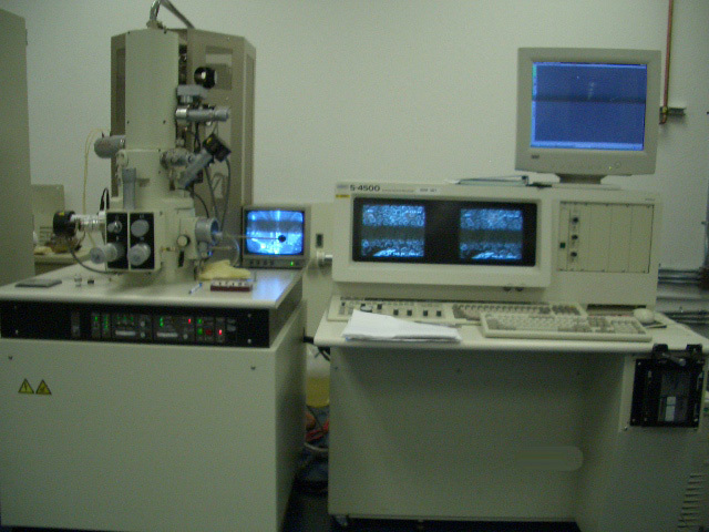
Used HITACHI S-4500 #9008633 for sale
URL successfully copied!
Tap to zoom


ID: 9008633
SEM Cold Field Emission System
Performance:
Secondary electron image resolution: 1.5 nm at 15kv
Magnification: 20x - 500kx
Electron optics:
Electron gun: cold field emmission source
Lens type: electromagnetic
Objective aperture: 4 position externally selectable
Stigmator: octopole electromagnetic
Scanning coil: 2-stage electromagnetic
Sample chamber
Size: type i
Airlock: prepumped, max sample size:50 mm diameter
Stage motion: 5 axis manual x/y: 25 mm, z: 3-28 mm, t: -5deg - +45 deg., r: 360 deg continuous
Draw-out door: max sample size:150 mm dia.
Size: type ii
Airlock: prepumped, max sample size:100 mm or 150 mm diameter
Stage motion: 5 axis manual
X/y: 100mm/50 mm, z: 3-33 mm, t: -5 deg - +60 deg., r: 360 deg continuous
Display system:
Image display: dual 12" monitors
Scanning mode: normal, reduced area , line scan, photo scan, spot position, split screen
Scanning speed: tv, 0.3, 2, 9, 25, 35, 100, 160 320 s/frame vsignal processing:real-time processing, auto-brightness and contrast control, dynamic stigmator, sutofocus, a vframe averaging, frame integration, contrast conversion,
Vacuum system:
Full automatic operation with pneumatic valve control
Ultimate vacuum: 10 (-7) pa in electron gun chamber, 10(-4) pa in specimen chamber
Ion pump: 60 l/s x1, 20 l/s x2
Speciment chamber: dp (570 l/s); turbo optional
Foreline: rotary pump x2
Accessories (optional)
Edx:at 30 deg. Take off angle
Digital image capturing: orion-6 software, optional
Chilled water circulator: 10 - 20 deg c, 1.0 -1.5 l/m for dp only
Installation
Ac: single phase ac 220 or 240 volt, 50/60 hz
Grounding: independent grounding 100 ohms or less.
HITACHI S-4500 is a scanning electron microscope (SEM) designed for advanced materials analysis and imaging. This machine is capable of achieving a high level of detail, making it ideal for analysis in fields such as nanotechnology and bioscience. HITACHI S 4500 has several key features that make it a powerful tool for research and development. Firstly, the microscope comes equipped with a field-emission electron source and an electrostatic lens system. This system provides ultra-high resolution imaging, as small as 0.4 nm for both secondary electron and backscatter electron imaging. Additionally, S-4500 operates with a variable pressure chamber, meaning it can adjust the electron beam and specimen chamber pressure to the ideal levels while keeping charging effects in check. This allows it to produce high quality images with fewer artifacts and more accurate surface features. Furthermore, the microscope is capable of advanced spectroscopy and modality imaging. It includes features such as energy dispersive spectroscopy, energy filtered imaging, X-ray mapping, Auger spectroscopy, and more. This allows it to utilize a wide range of imaging and spectroscopy treatments, making it a great asset for materials science and research. Finally, S 4500 offers a host of accessories for improved image quality and higher resolution scanning. Options include BSE detectors, a range of objective lenses, a secondary electron backscatter detector, and a range of accelerating voltage filters. With all these components in place, the user can easily fine-tune the equipment to their needs. Thanks to its features, HITACHI S-4500 is a powerful tool for research and materials analysis. It is able to detect and image even the smallest of features with great accuracy, while the range of spectroscopy and modality imaging makes it an incredibly versatile machine. With the extra accessories, HITACHI S 4500 can be adjusted for even higher performance, making it an even more useful and reliable tool.
There are no reviews yet