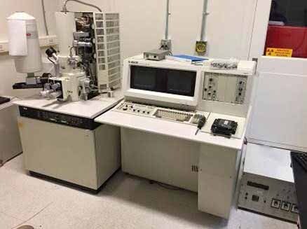
Used HITACHI S-4500 #9169489 for sale
It looks like this item has already been sold. Check similar products below or contact us and our experienced team will find it for you.
Tap to zoom


Sold
ID: 9169489
Scanning electron microscope (SEM)
Specifications:
Photo CRT:
Ultra high resolution type
Recording area: 120 x 90 mm
Scanning mode:
Normal scan
Split screen / Dual magnification
Line scan
Position setting
Spot
AAF (Analysis area finder)
SAA (Selected area analysis)
Oblique mode
Scanning speed:
0.3, 2, 10, 20 S / Frame for viewing mode
TV, 40,80, 160 S / Frame for photo recording mode
Signal processing / Operation mode:
Automatic brightness & contrast control
Gamma control
Dynamic stigmator monitor
Auto focus
Automatic stigmator
Electrical image shift: ± 20 µm With WD 20 mm
Vacuum system:
Vacuum sequence: Full-auto pneumatic valve system
Ultimate vacuum:
7 x 10⁻⁴ Pa (Specimen chamber)
1 x 10⁻⁷ Pa (Electron gun chamber)
Vacuum pumps:
Electron optics: (3) Ion pumps
Specimen chamber:
(1) Diffusion pump
(2) Rotary pumps
Compressor
Ion pumps:
IP1= 0.1 x 10⁻⁷
IP2= 0.5 x 10⁻⁷
IP3= 6 x 10⁻⁷
Accessories:
THERMO-SCIENTIFIC EDS Tank
EDS Computer / Controller: No
GW Microchannel:
Control
Detector: No
GW Infrared: Camera control and camera on system
Control panel on SEM
Small manual stage
Performance:
Resolution: 1.5 nm
Accelerating voltage: 30 kV
Working distance: 7 mm
Magnification:
High magnification mode: 50x to 500,000x
Low magnification mode: 20x to 1,000x
Electron optics:
Electron gun: Cold-cathode field emission electron gun
Emission extracting voltage (Vext): 0 to 6.5 kV
Accelerating voltage (Vacc): 0.5 to 30 kV (Variable in 100 V step)
Lens system: 3-Stage electromagnetic lens
Objective aperture:
Movable aperture ((4) Opening selectable / Alignable outside column)
Self-cleaning type thin aperture
Stigmator: Electromagnetic type
Scanning coil: 2-Stage electromagnetic type
Display unit:
CRT: (2) 12" Display monitors including memory unit.
HITACHI S-4500 is a scanning electron microscope (SEM) specially designed for imaging the surface of a wide range of samples, from large specimens to small microstructures. With a maximum magnification of 270,000x and an ultra-high resolution of 0.7 nm, HITACHI S 4500 is capable of revealing the vast amount of detail contained within a single sample. S-4500 utilises a semi-apochromatic objective lens, consisting of two hemisphere bubble lenses and a projection lens, to focus the high energy electron beam to a fine point. The beam is scanned across the sample in a raster pattern and electrons that have been reflected from the sample surface are collected in an electron detector. These electrons are then converted into a digital image which is used to construct a 3D representation of the sample surface. S 4500 also features an automated integrated measurement and imaging function, allowing for measurements to be taken directly from the generated images. This allows for the quantitative analysis of samples including physical measurements and electrical characterisations. These measurements are crucial for a wide range of industries and research applications. HITACHI S-4500 has a vacuum environmental chamber, allowing it to be used with samples that need to be examined under vacuum conditions. This is especially important for the examination of samples such as biological tissue and semiconductor devices. The SEM also contains an automated specimen exchange system that enables samples to be changed quickly and efficiently without breaking vacuum. HITACHI S 4500 produces highly detailed images that enable scientists and engineers to study the surface of any given sample in intricate detail. This in turn allows them to analyse and identify defects, analyse materials composition and gain greater insight into the micro-structures of various components and products. As such, S-4500 from HITACHI is a valuable tool for many industries, from automotive engineering and microelectronics to materials science and bio-imaging.
There are no reviews yet