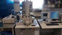
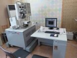
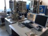
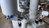
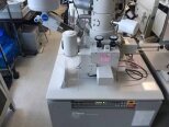
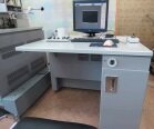
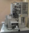
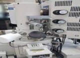
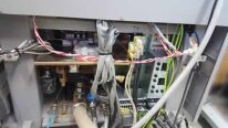
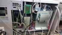
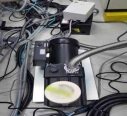
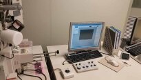
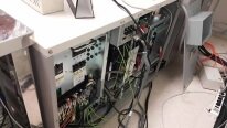
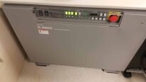
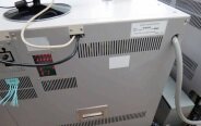
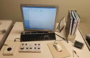
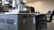
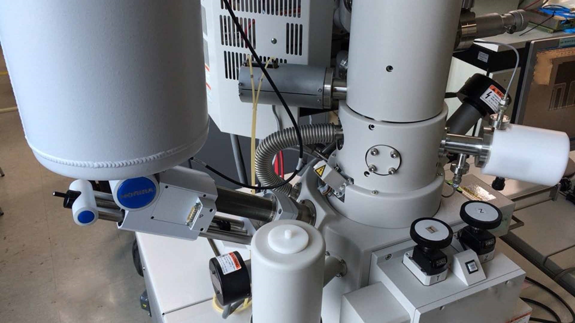
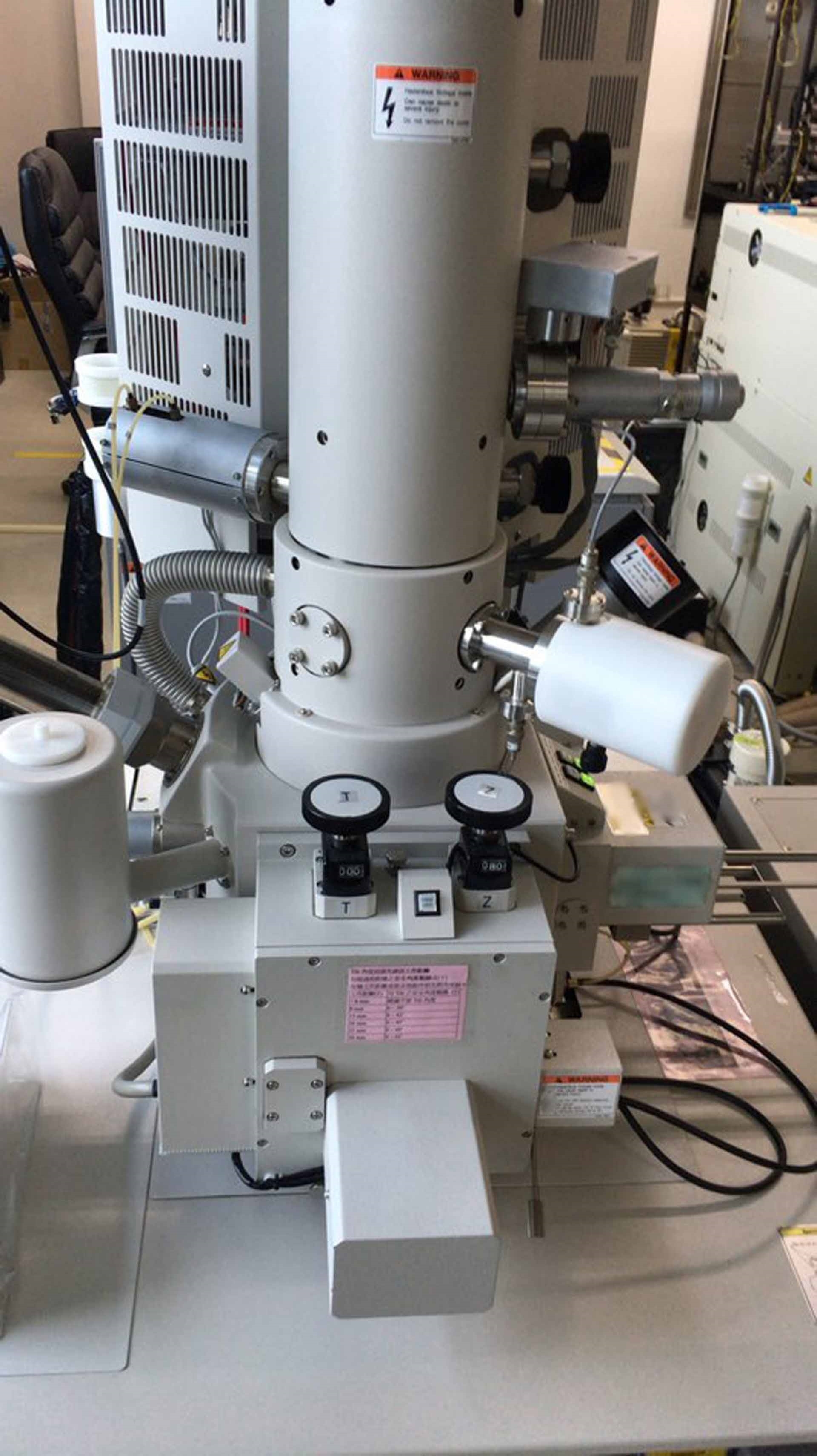
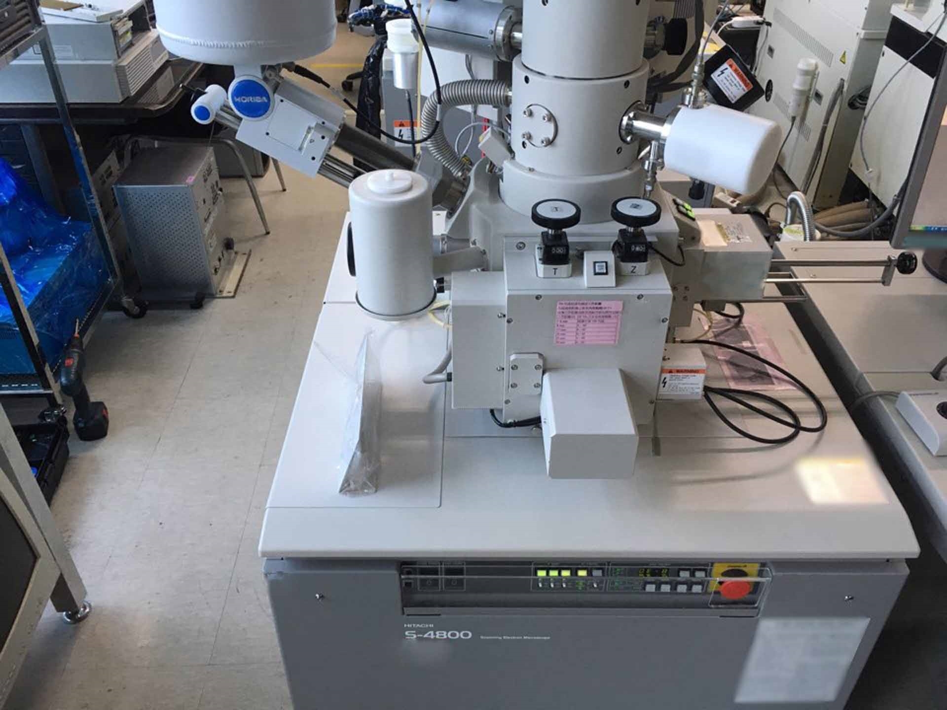
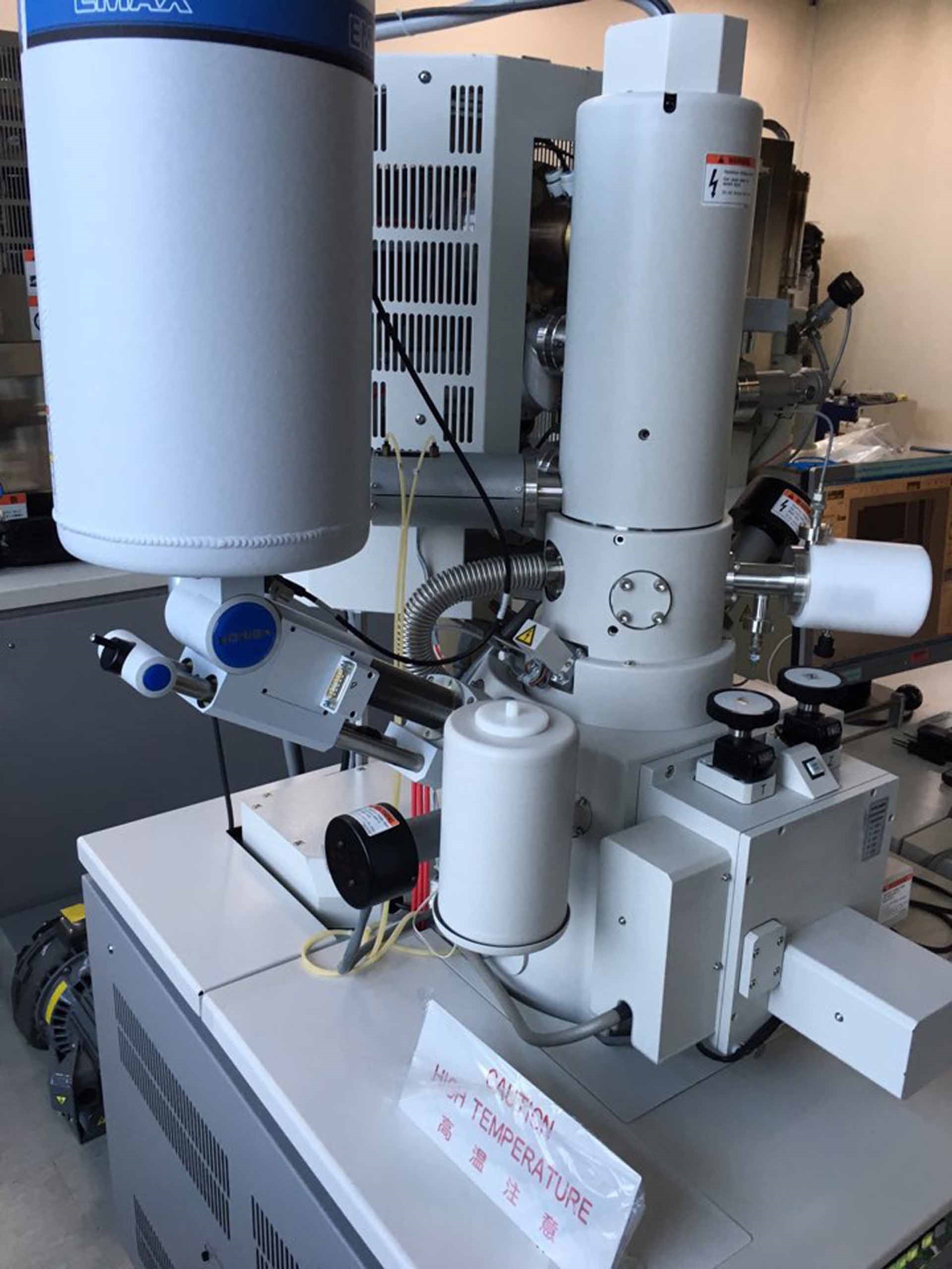
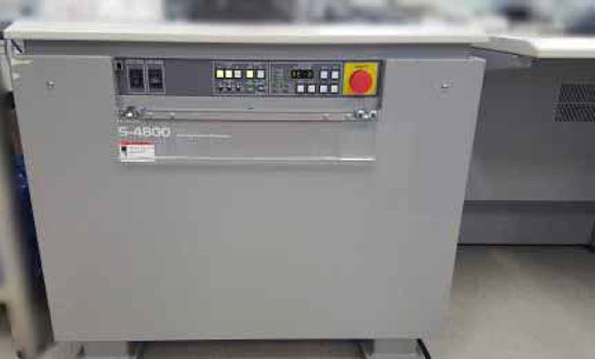
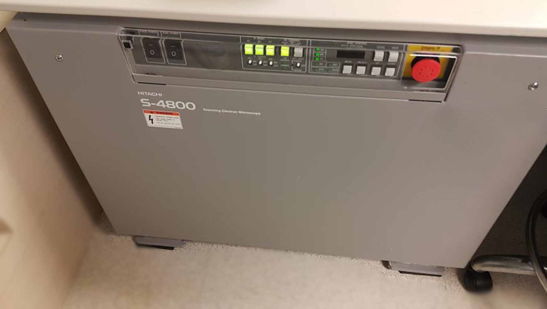
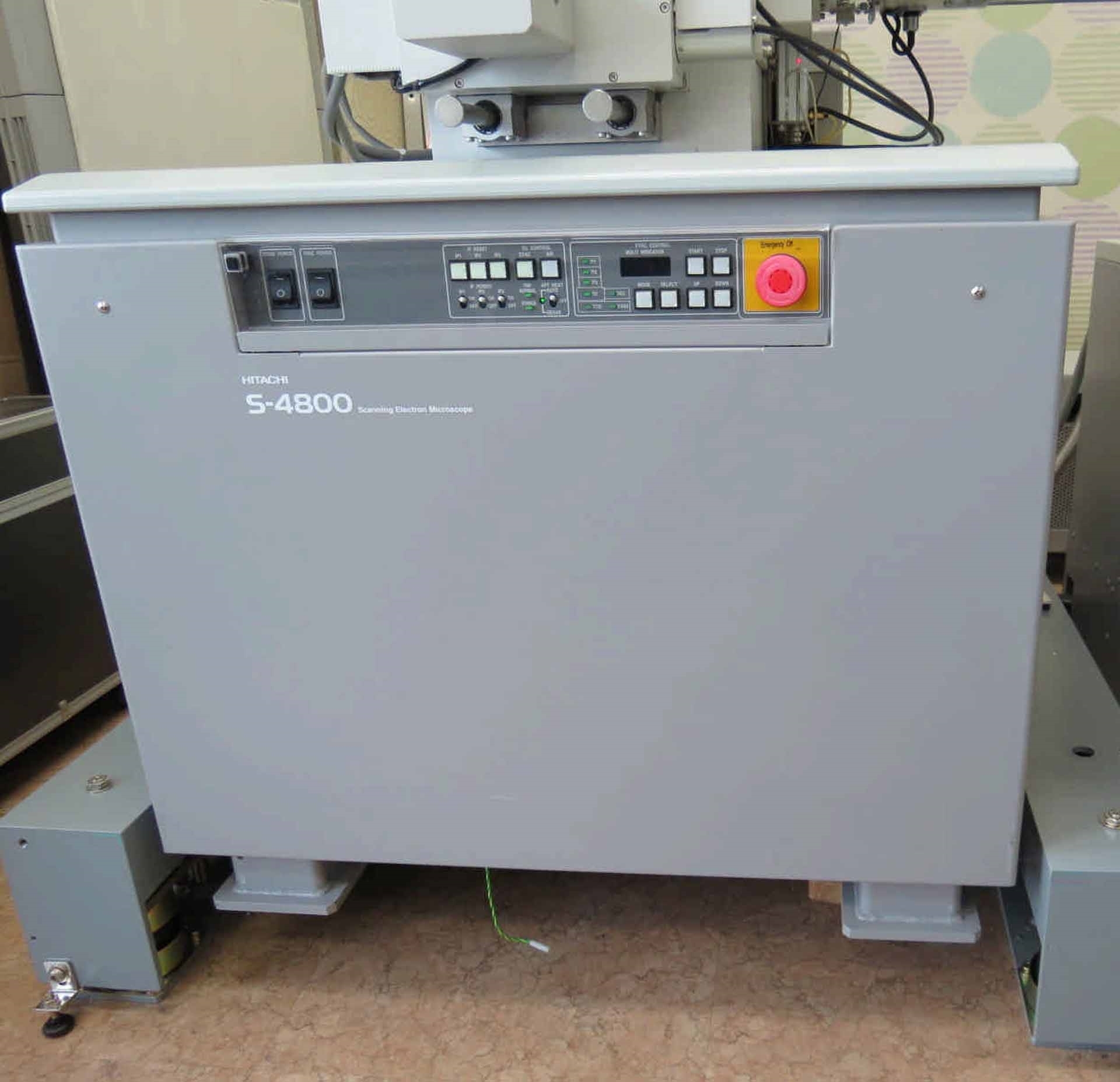
Used HITACHI S-4800 #9209753 for sale
URL successfully copied!
Tap to zoom






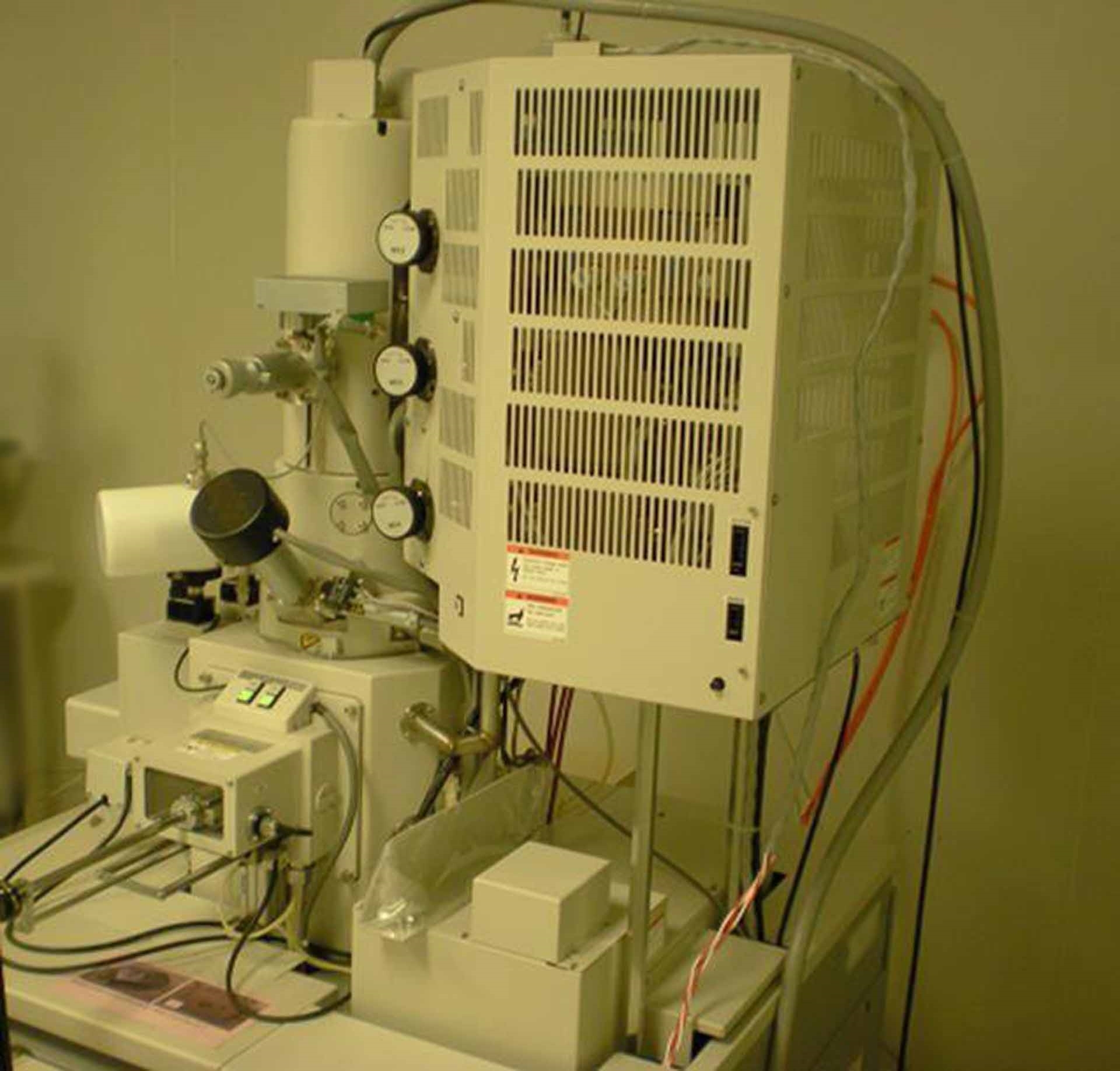

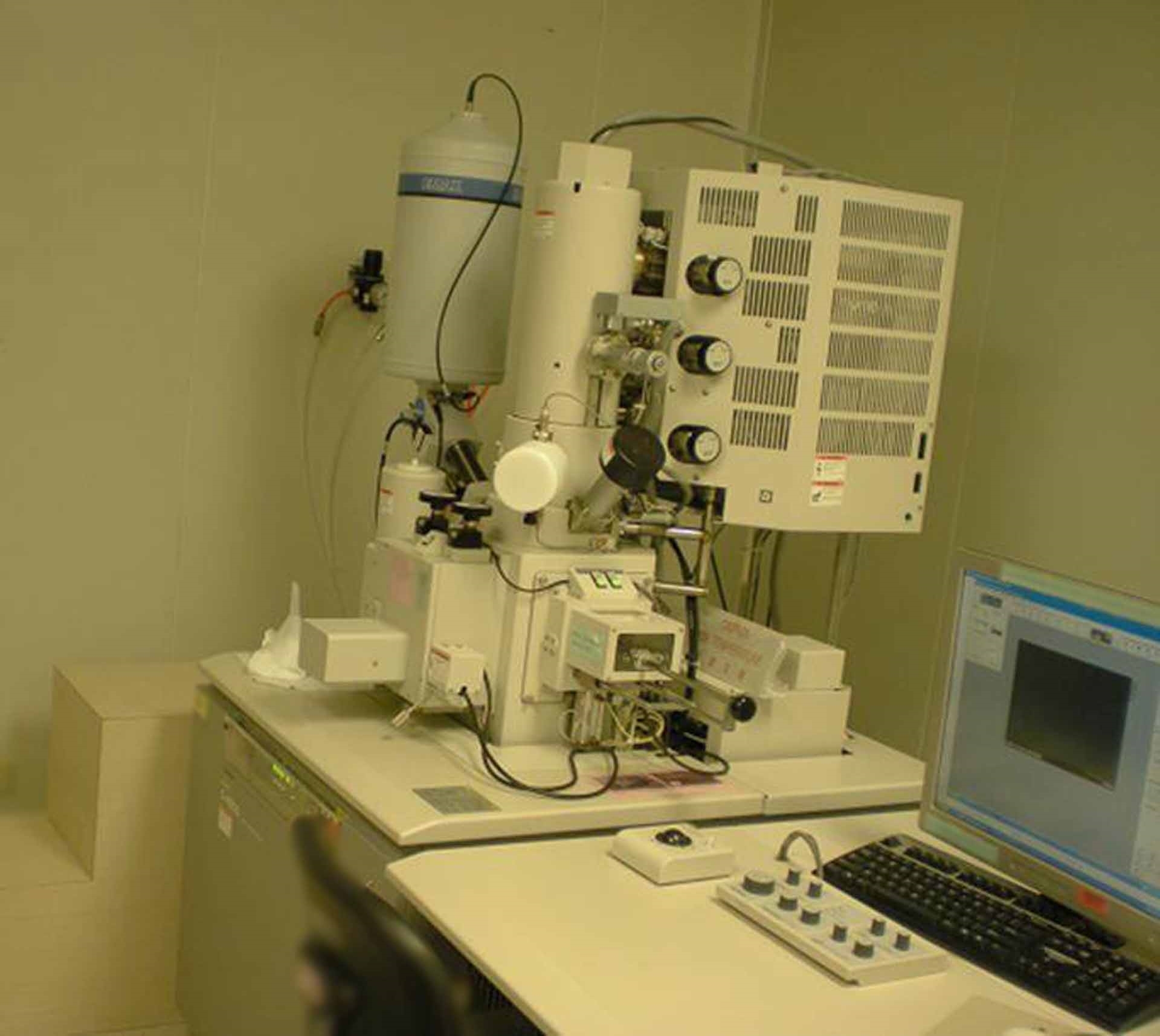

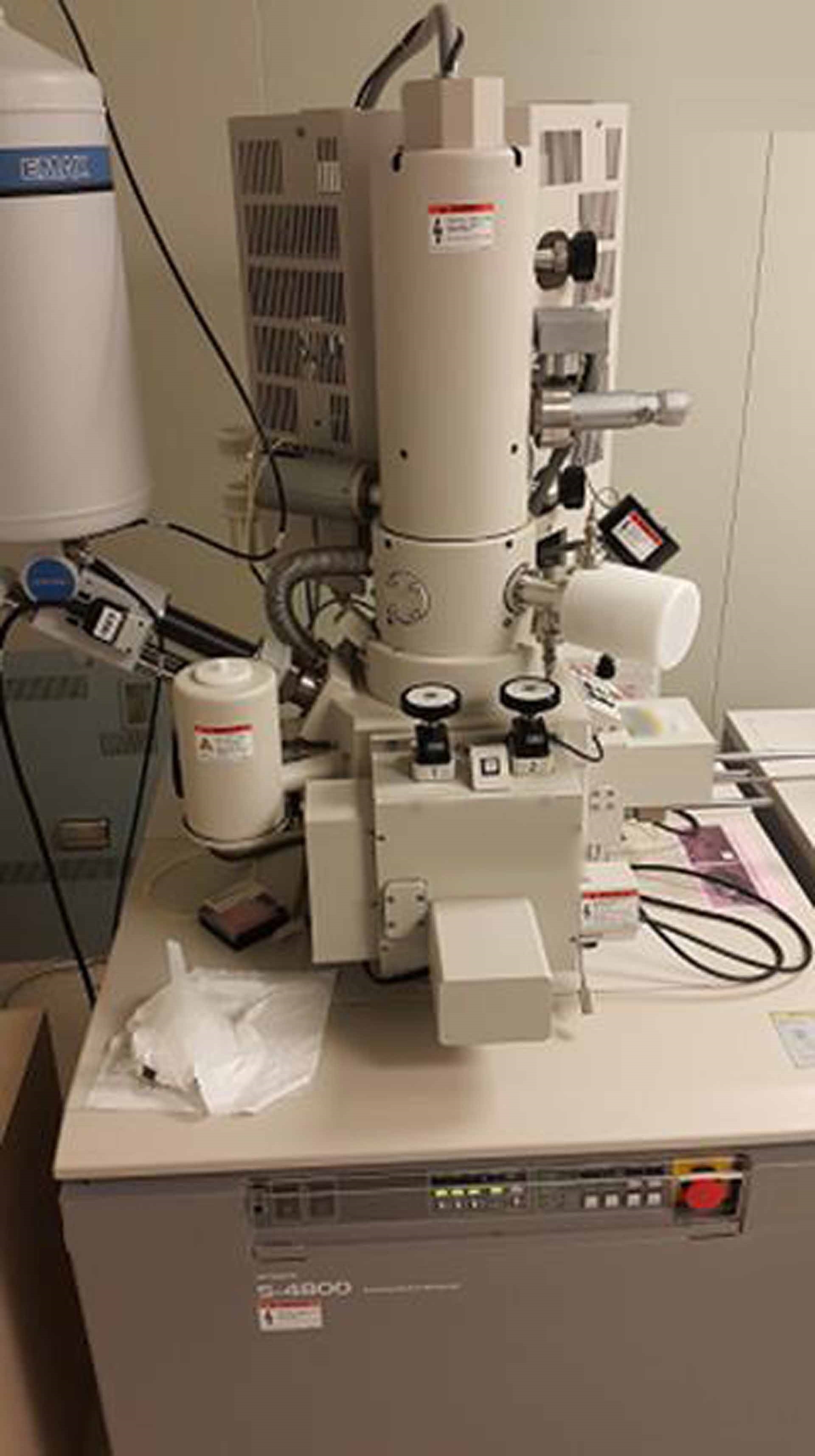

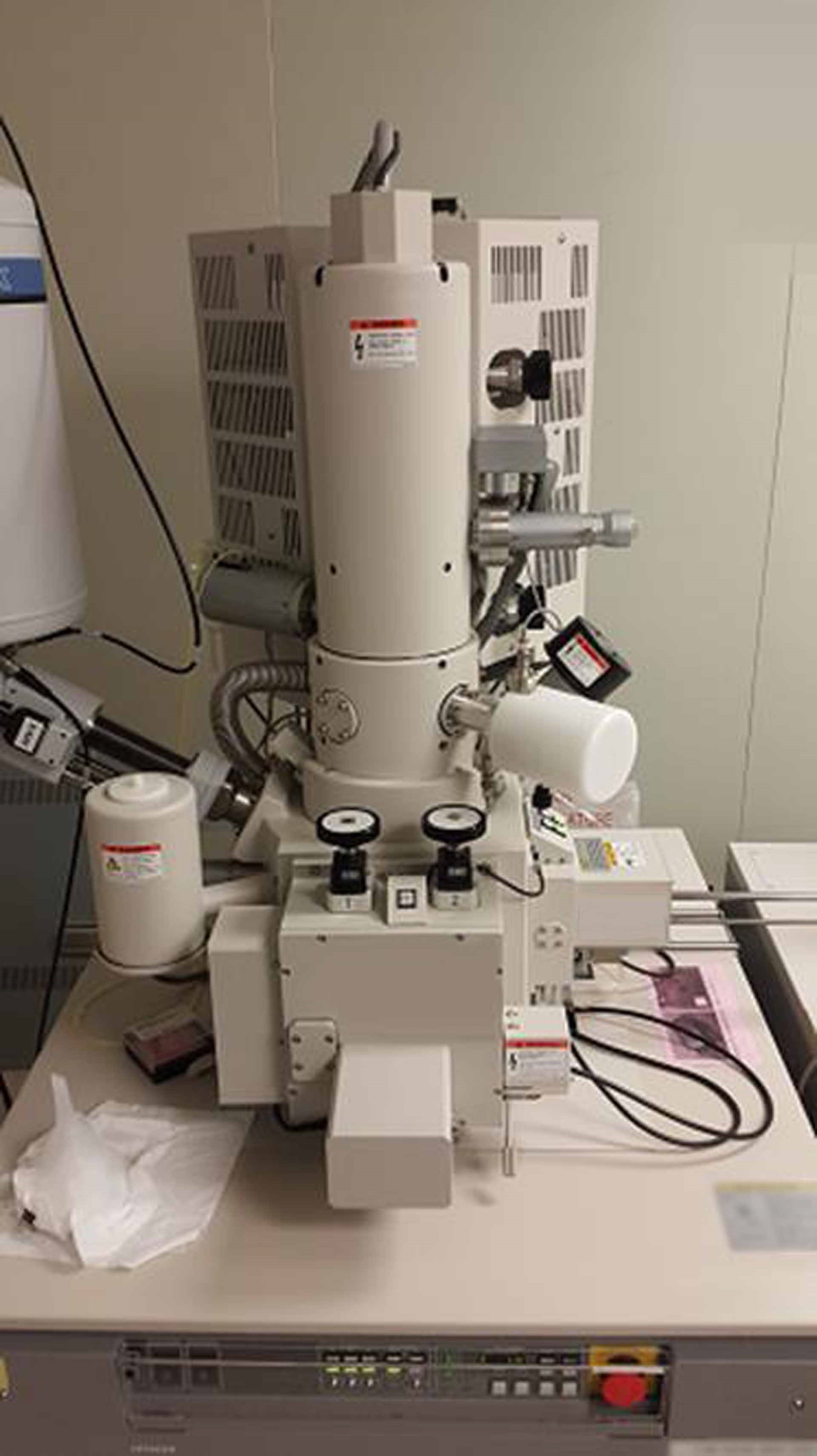









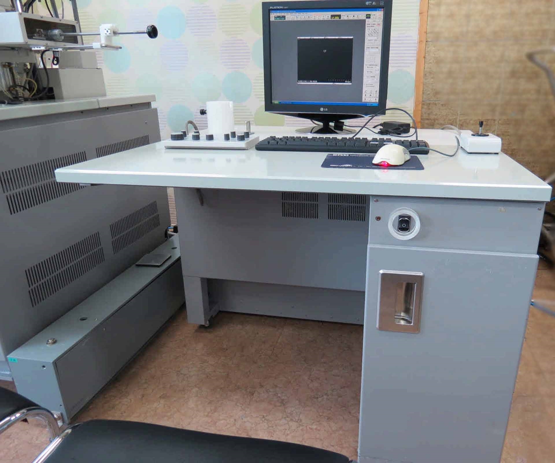

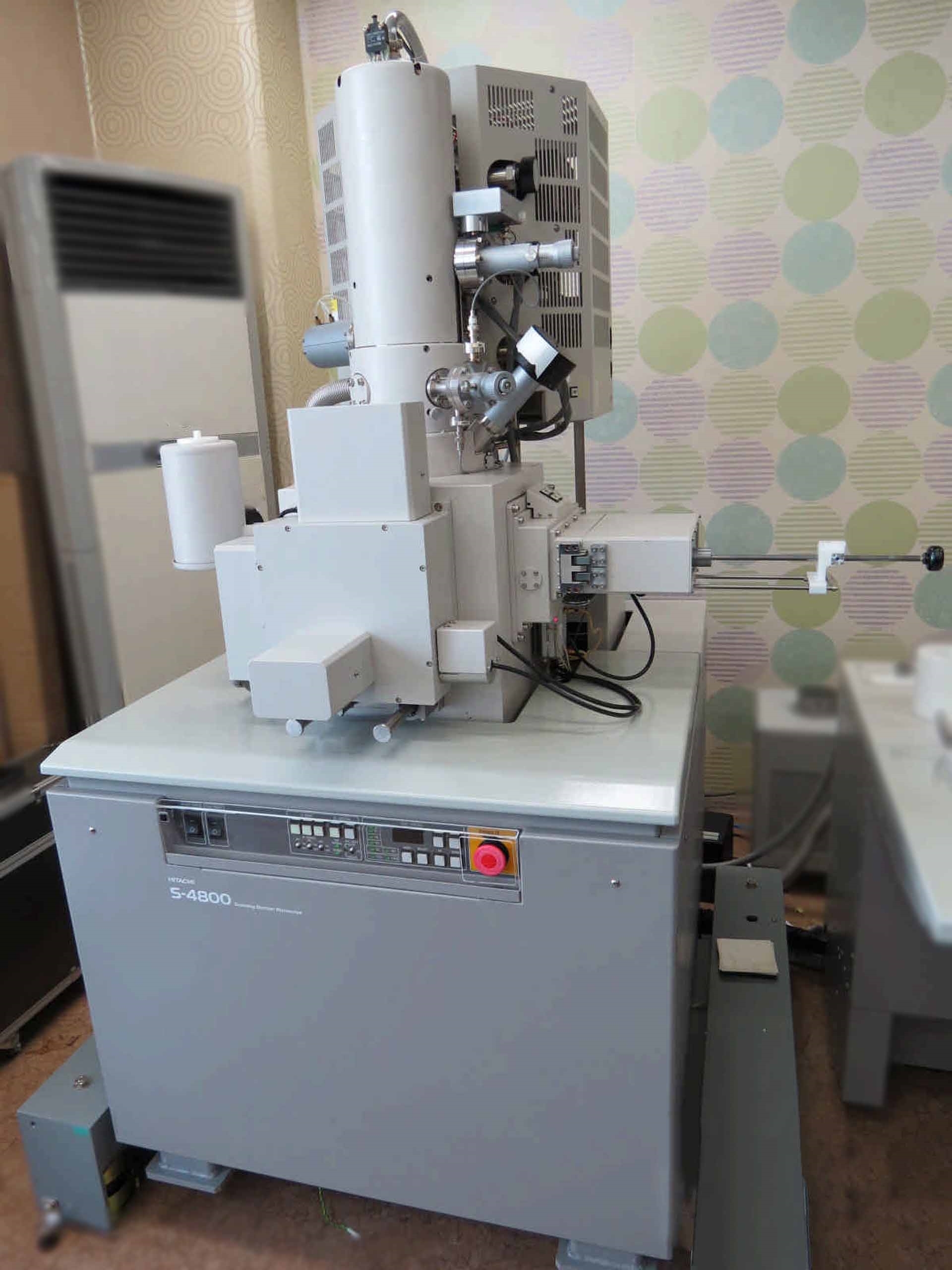

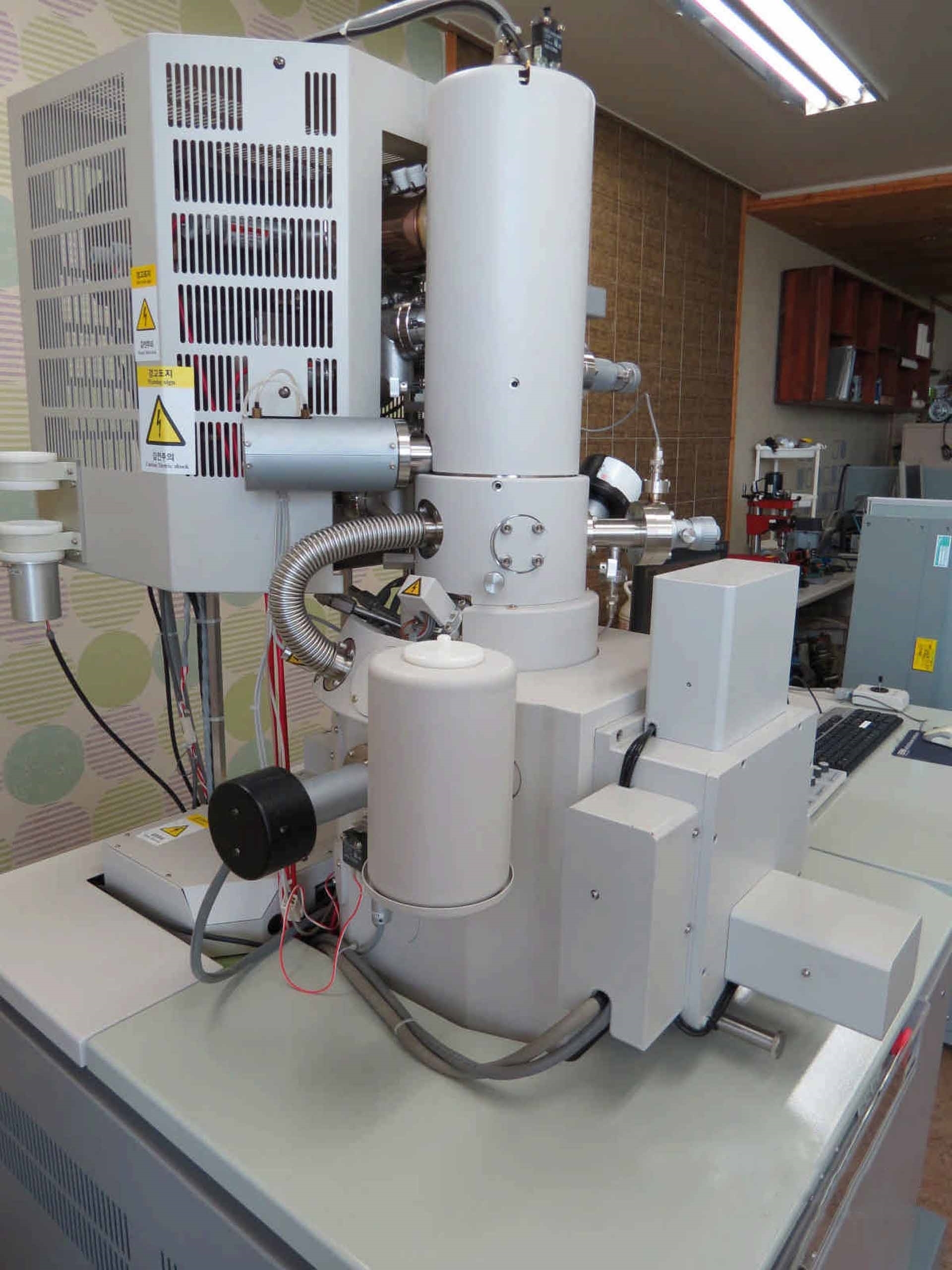

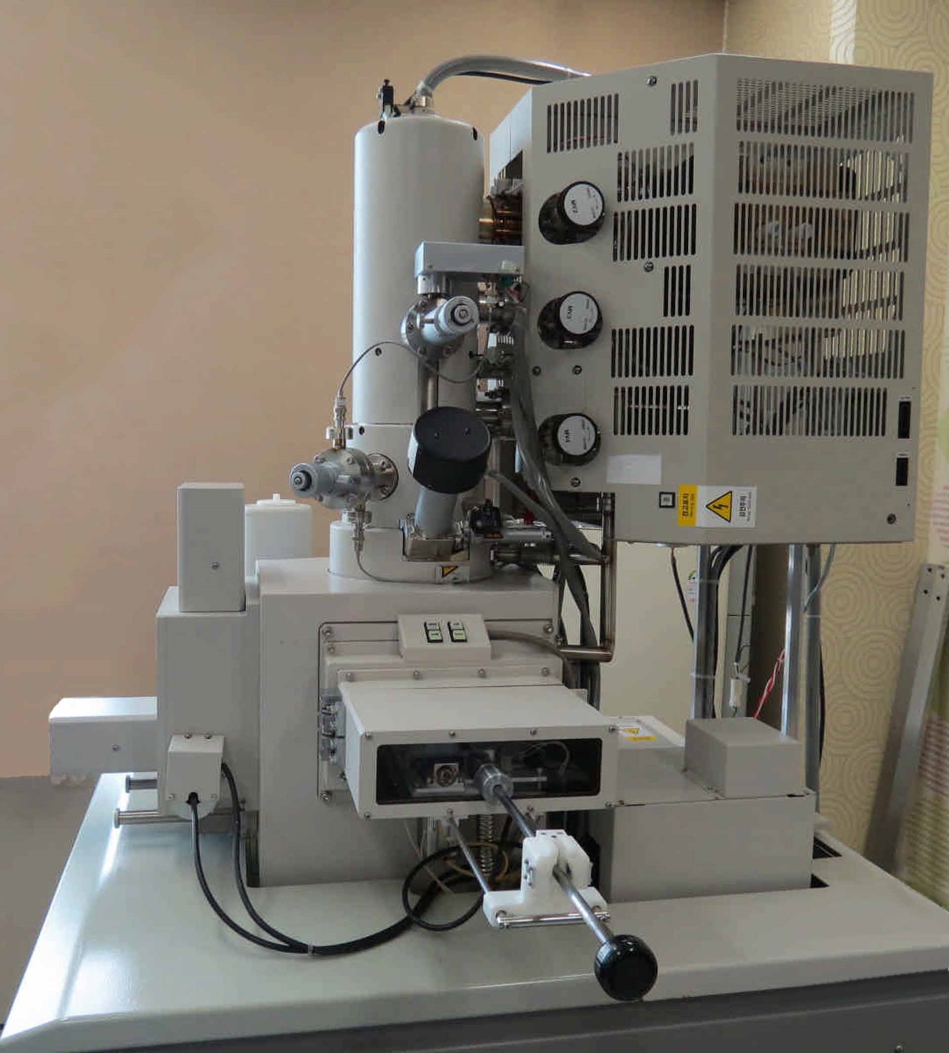









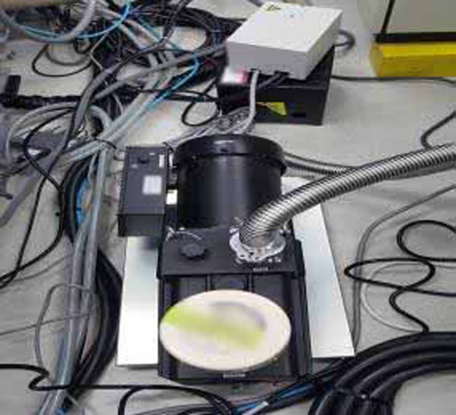
































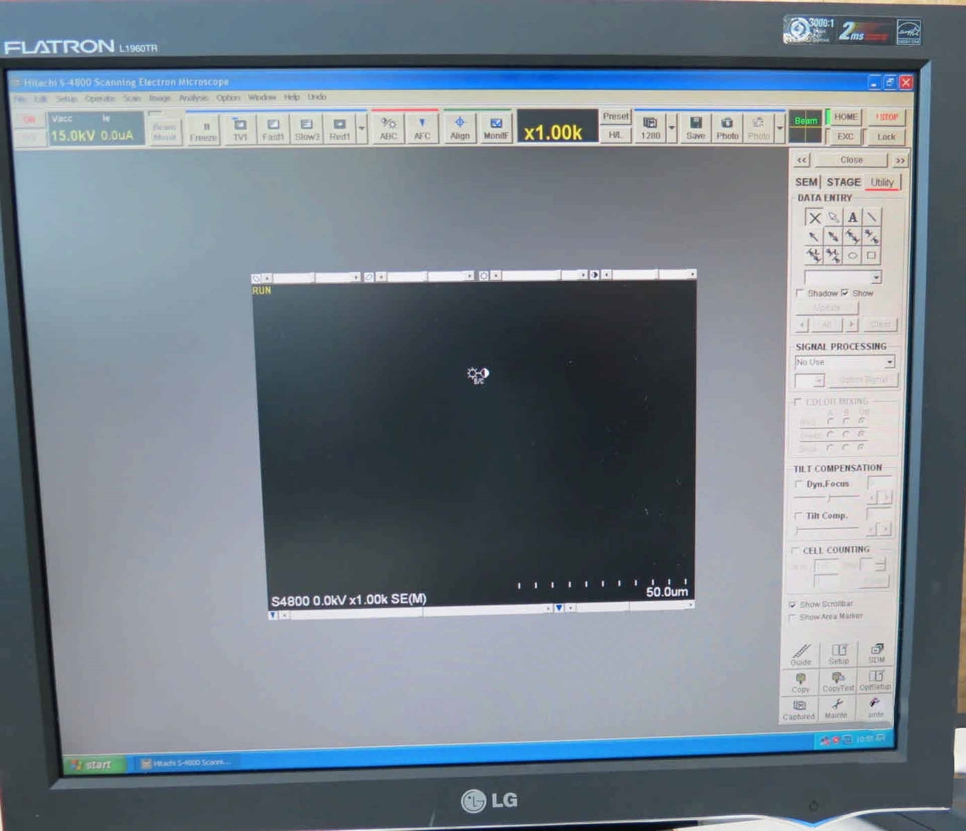


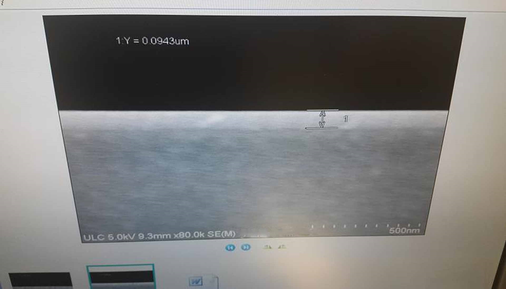

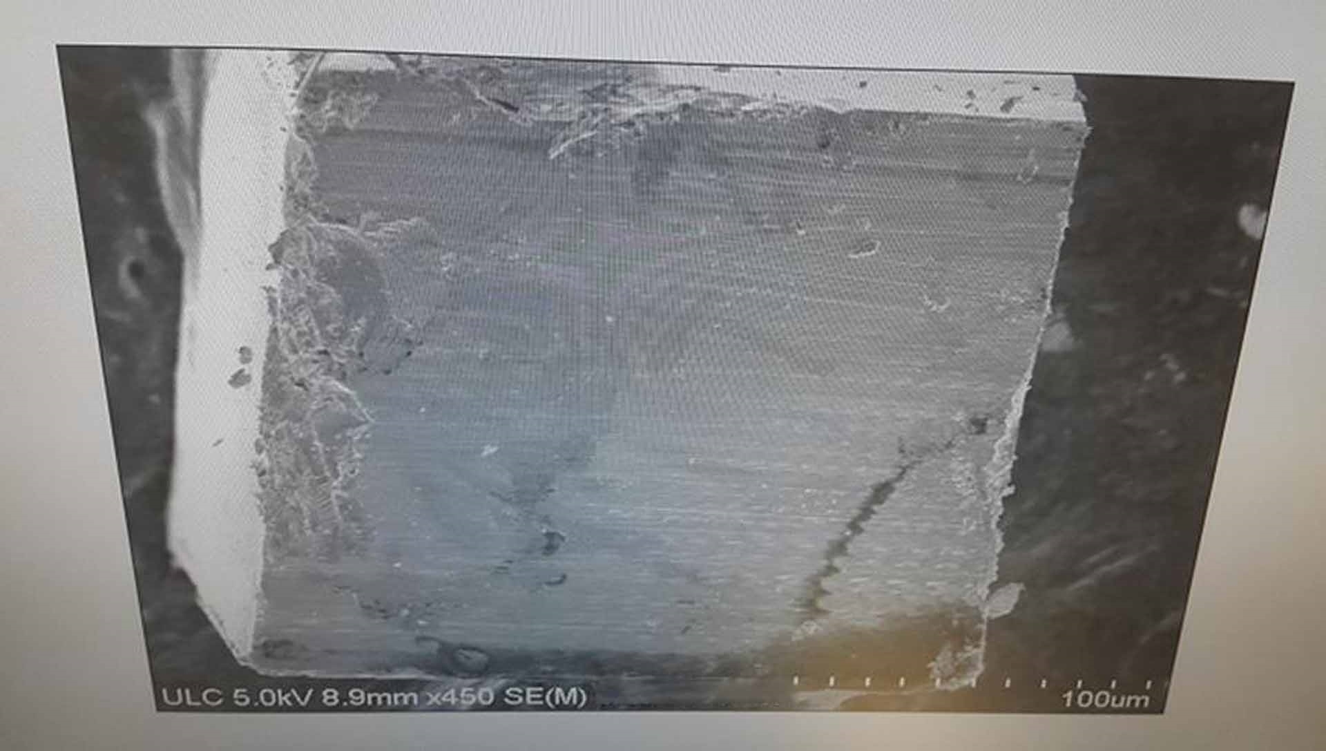

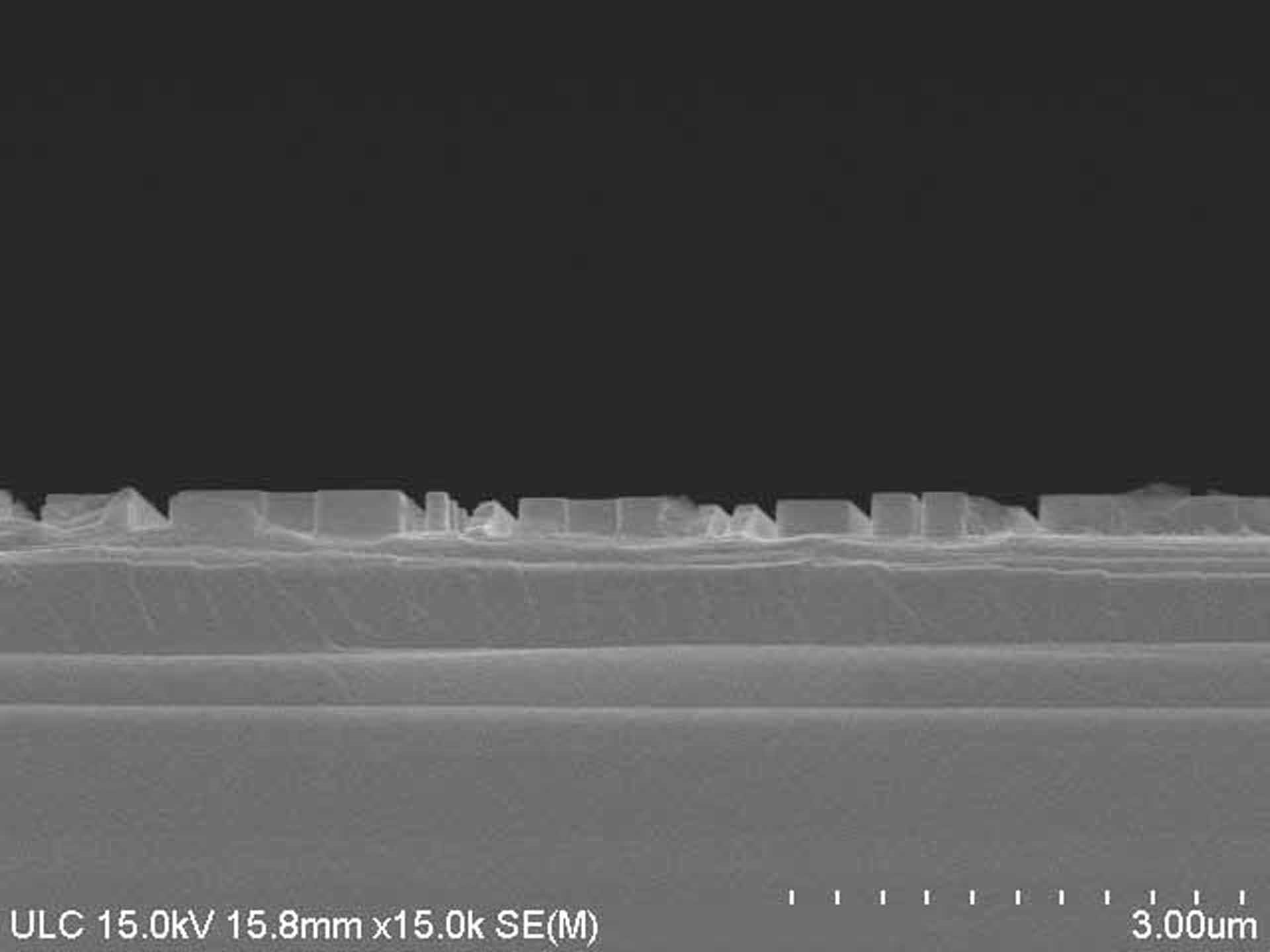

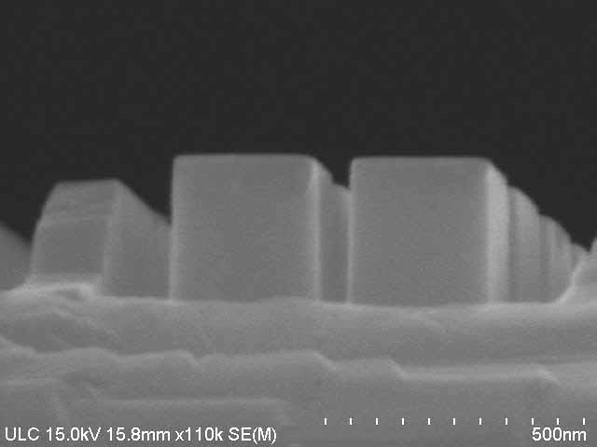

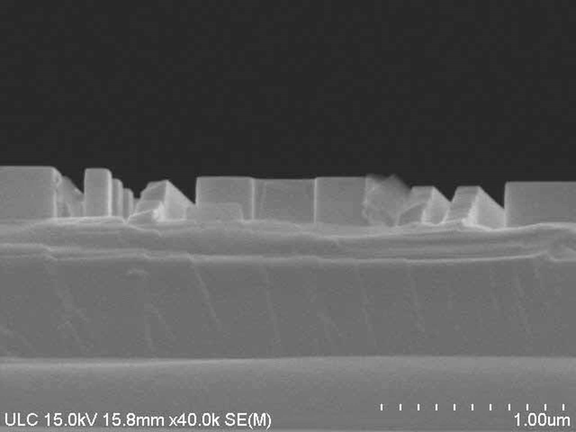

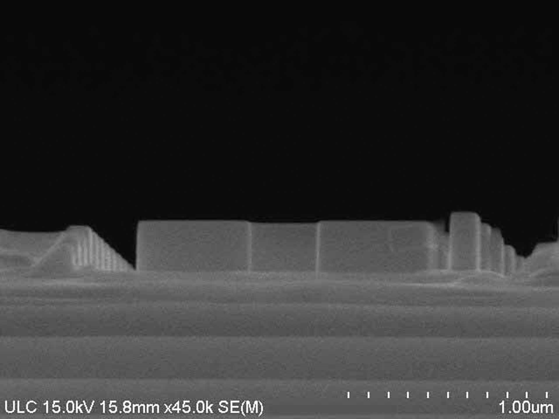













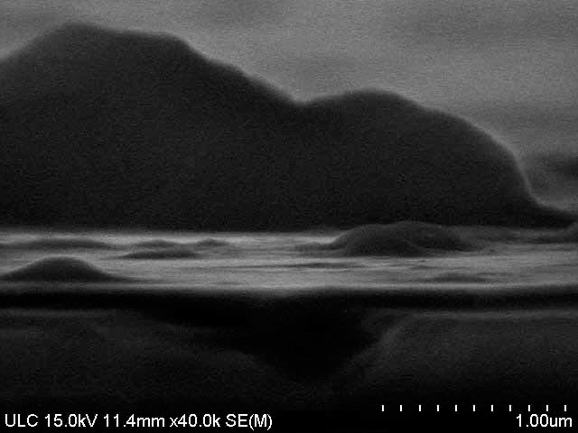

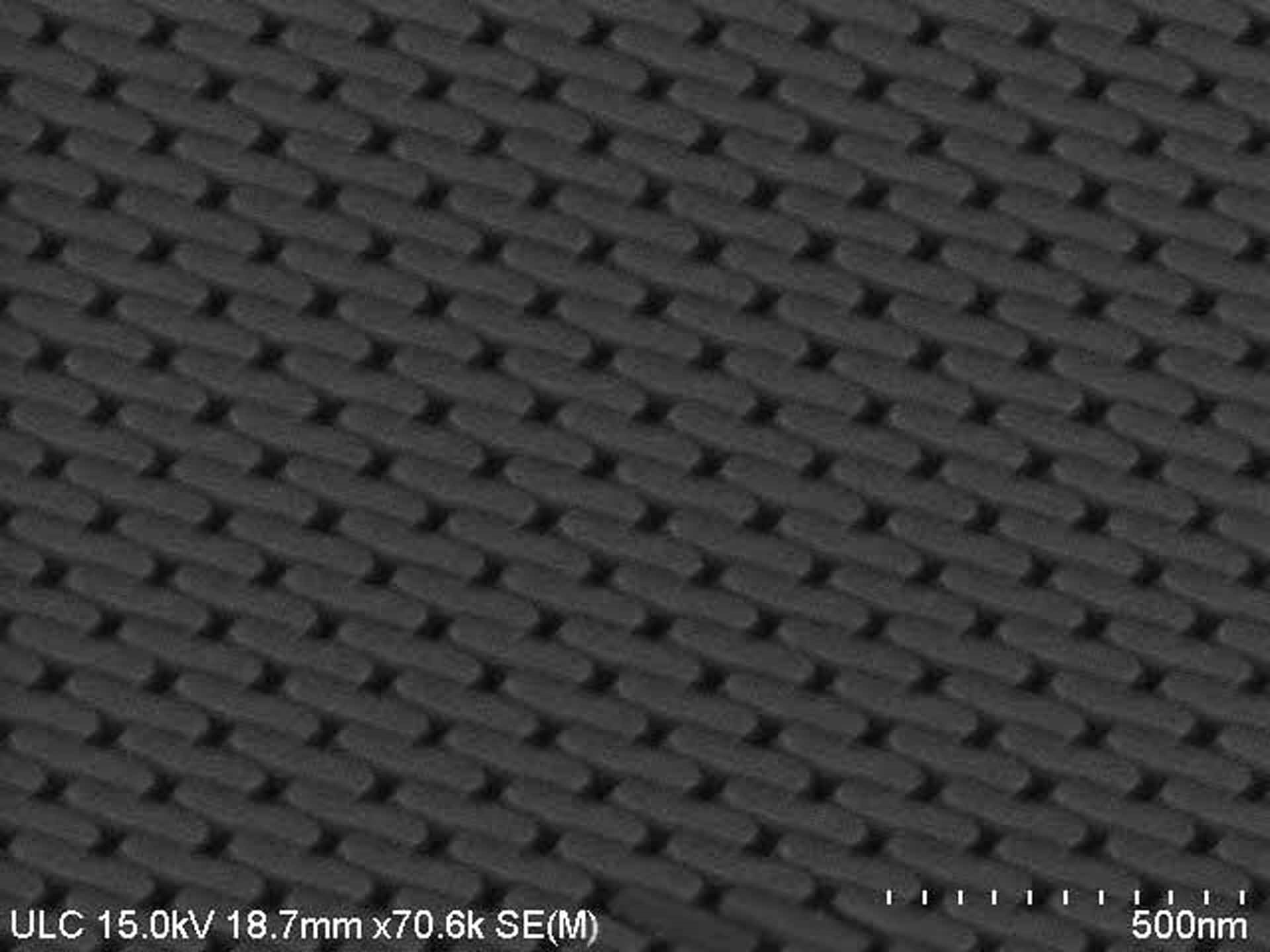

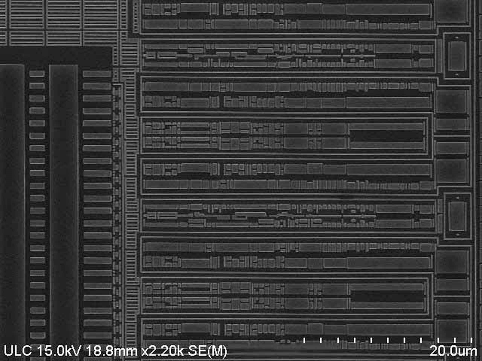

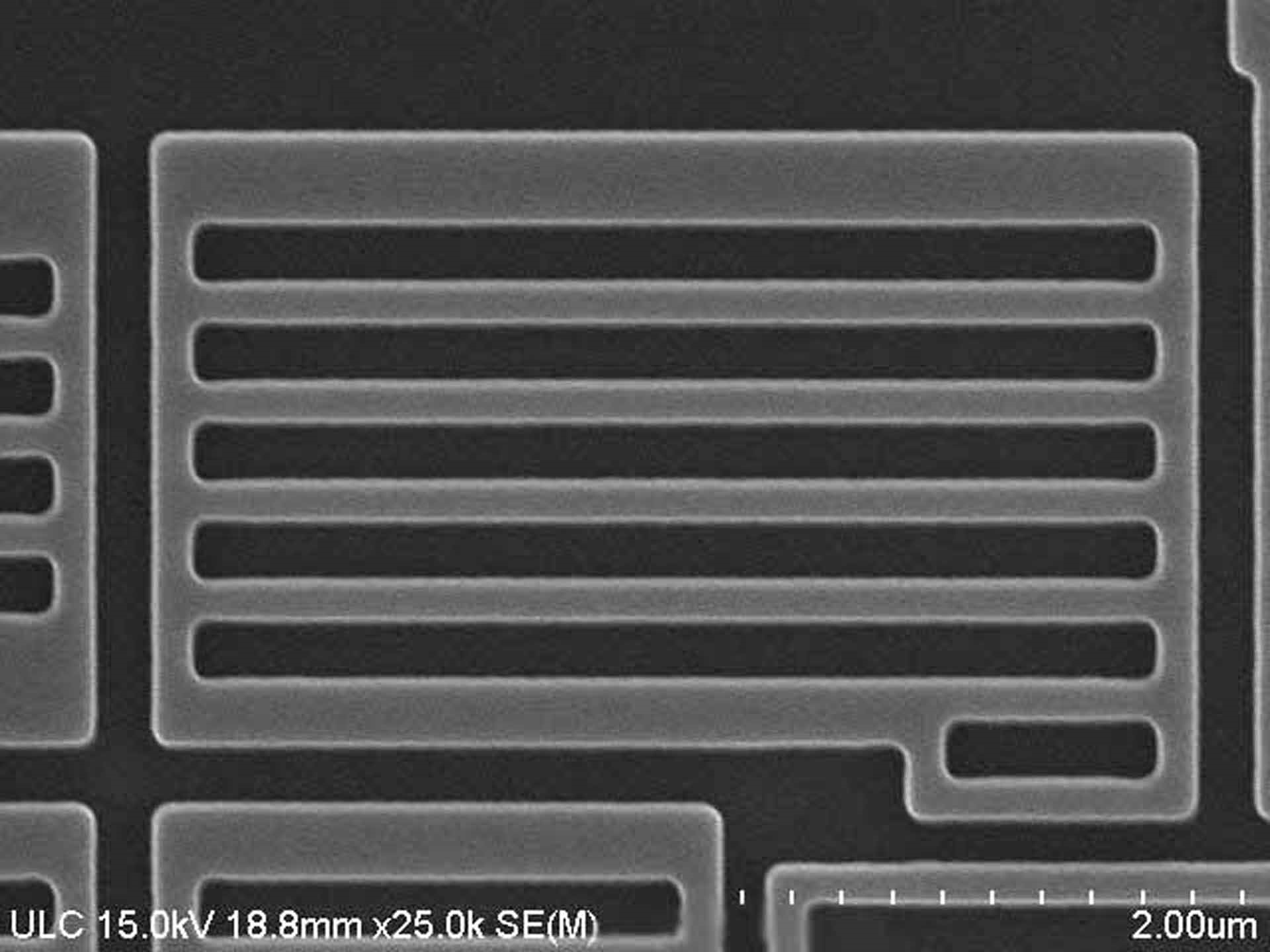

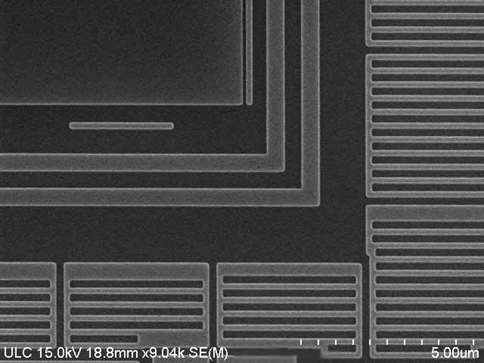

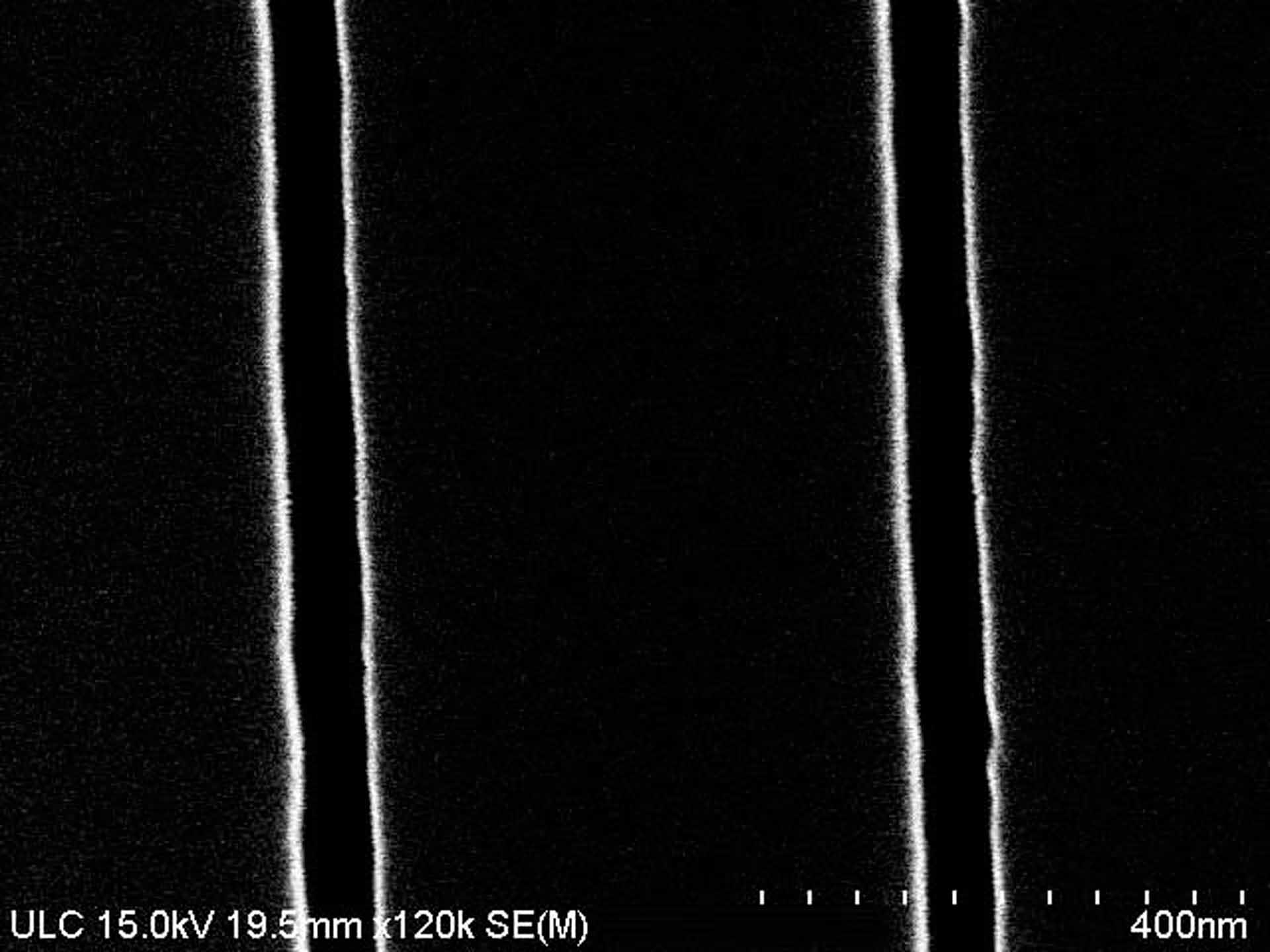

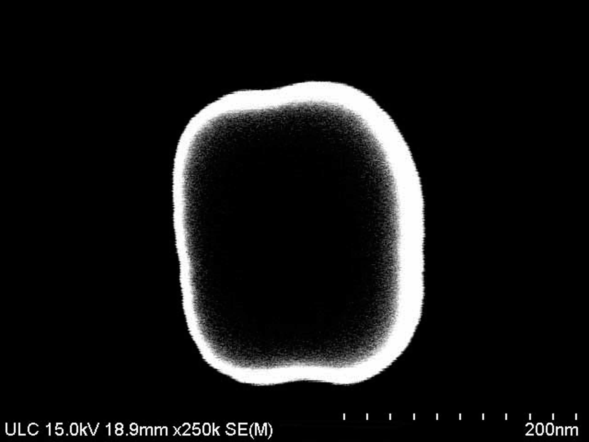

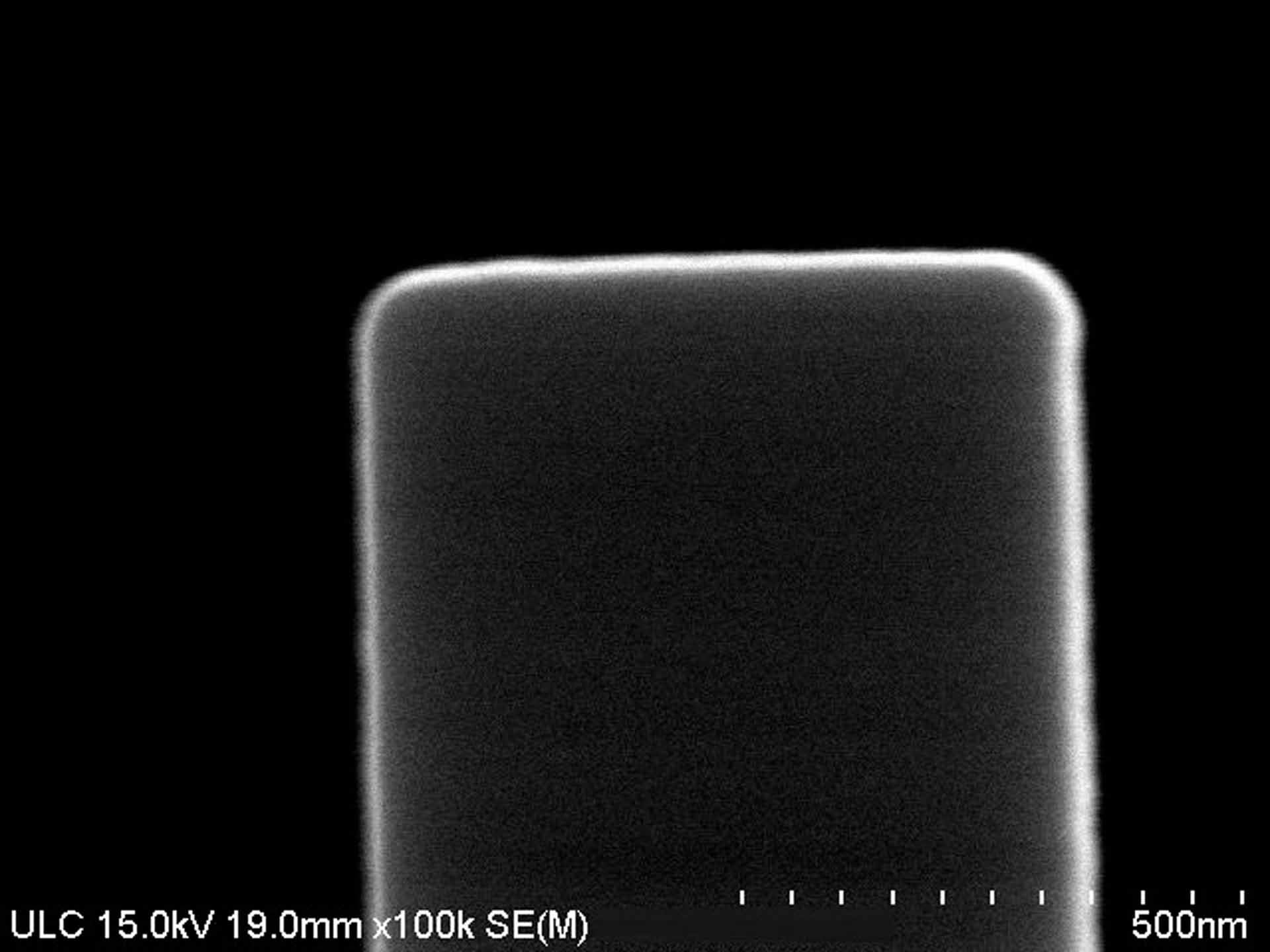

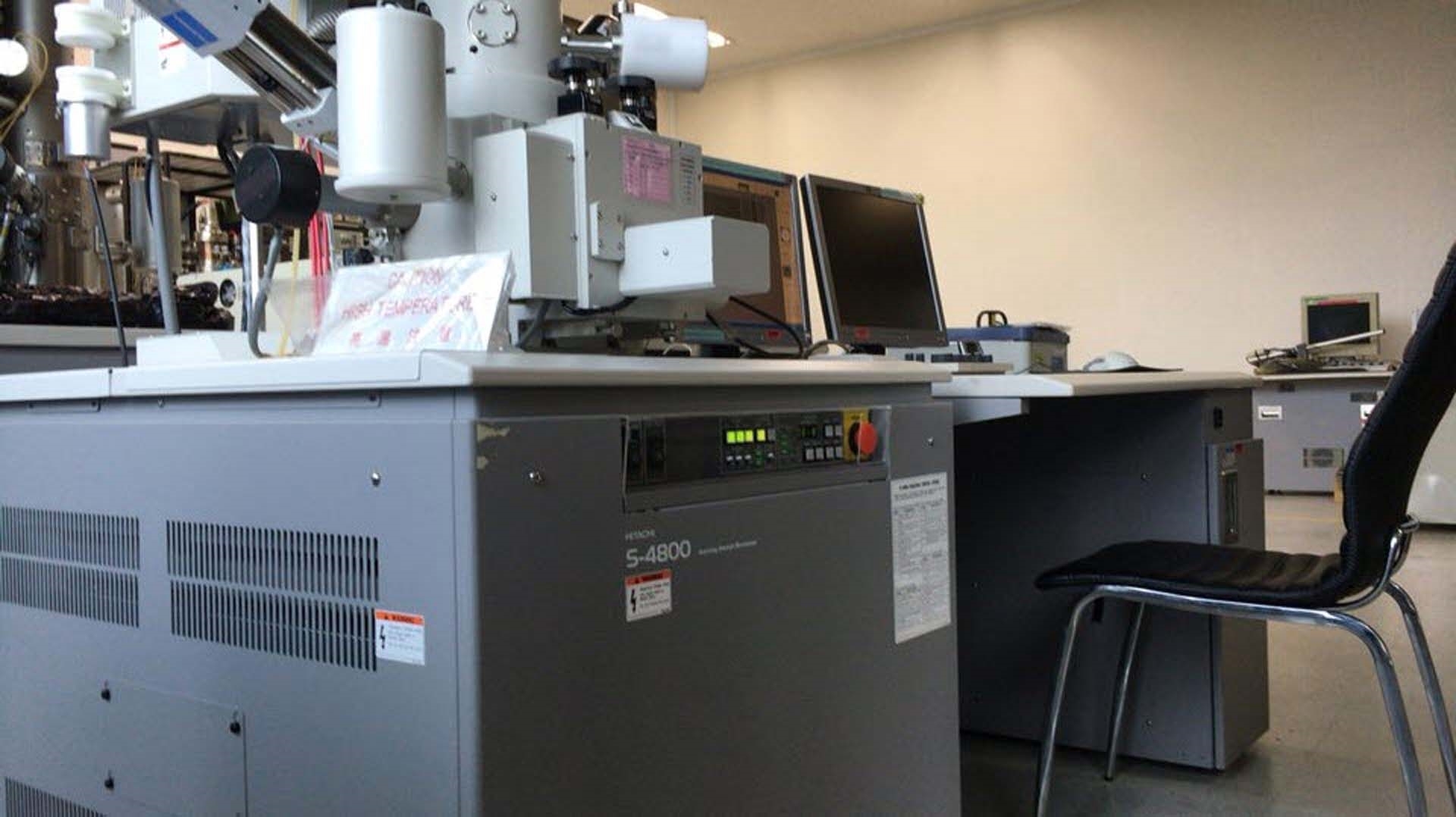

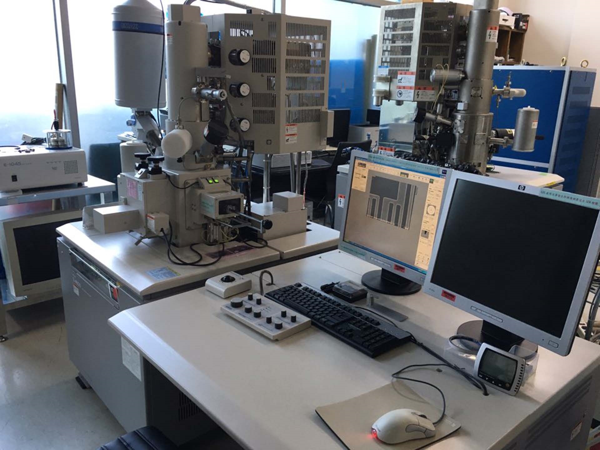

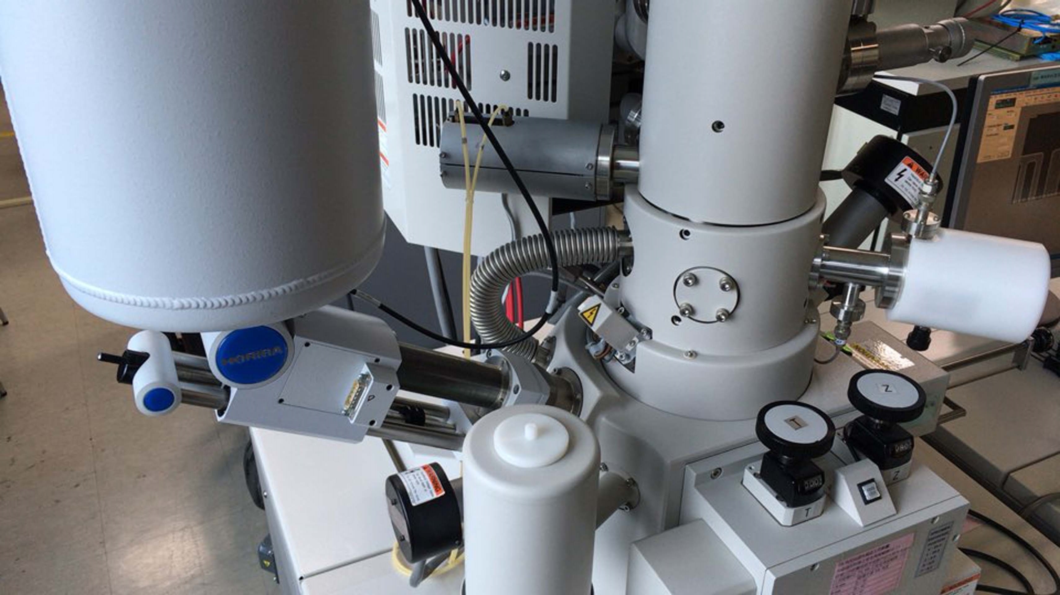


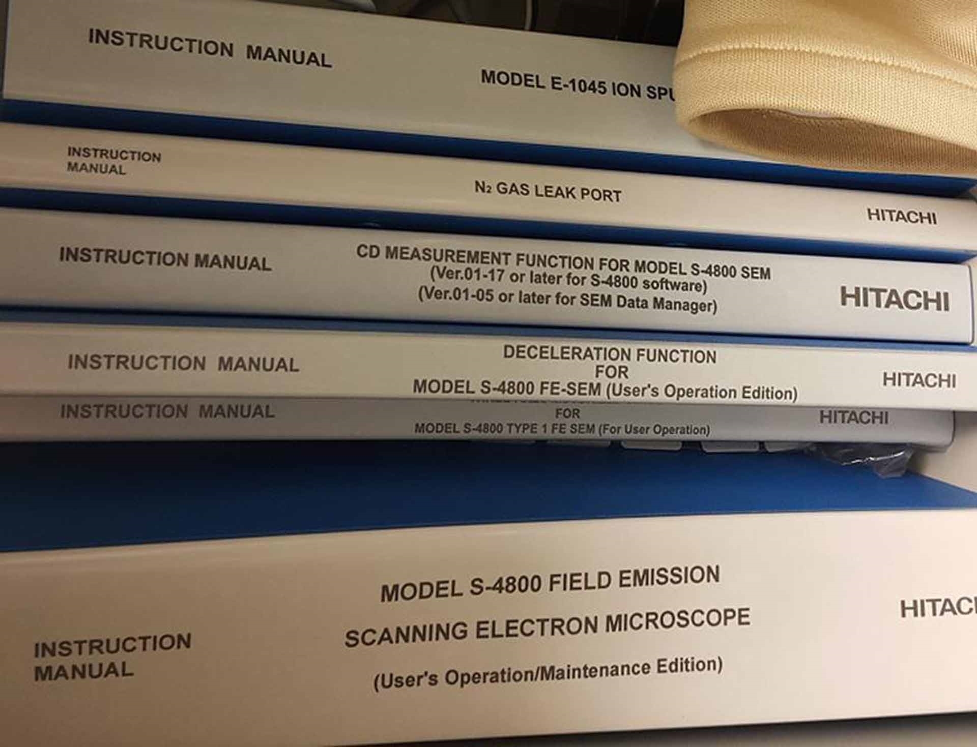







ID: 9209753
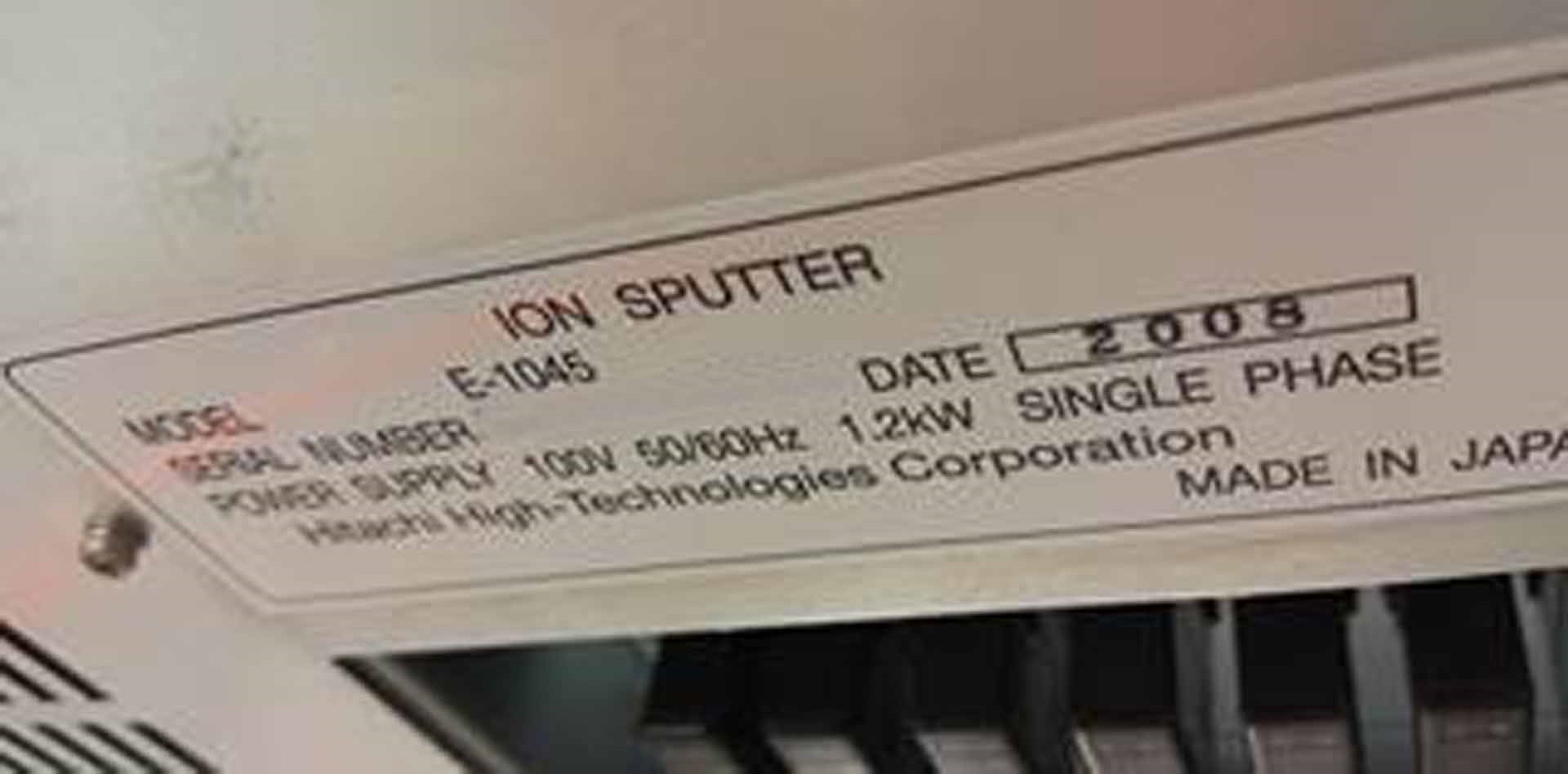
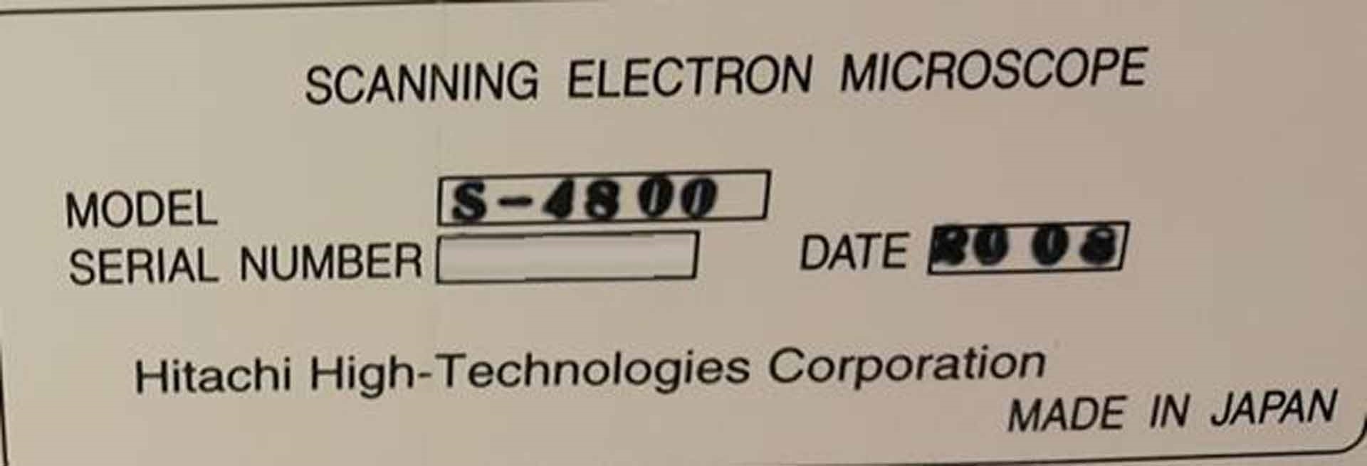
Vintage: 2008
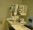
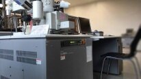
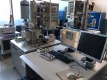
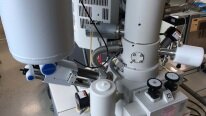
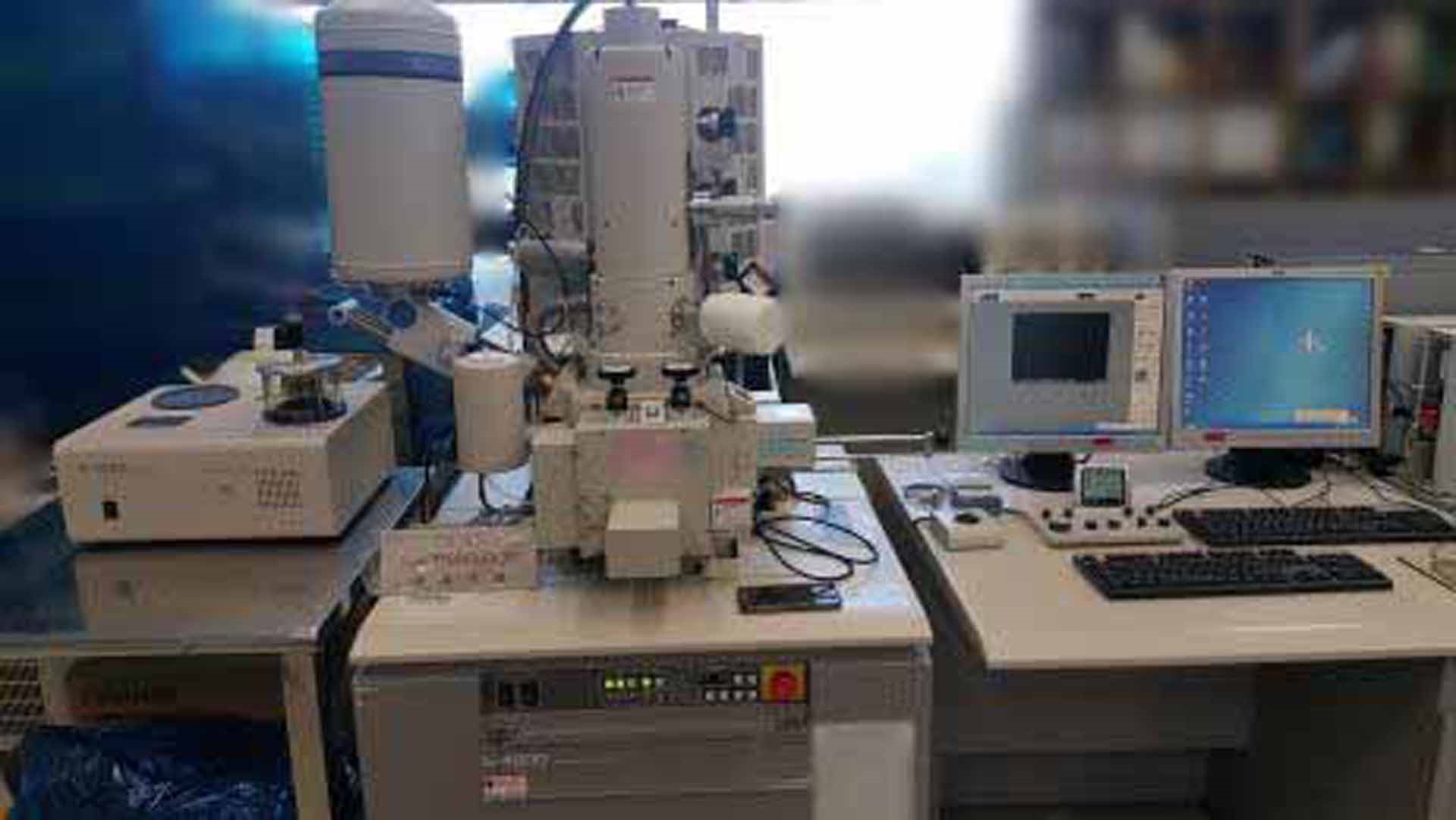
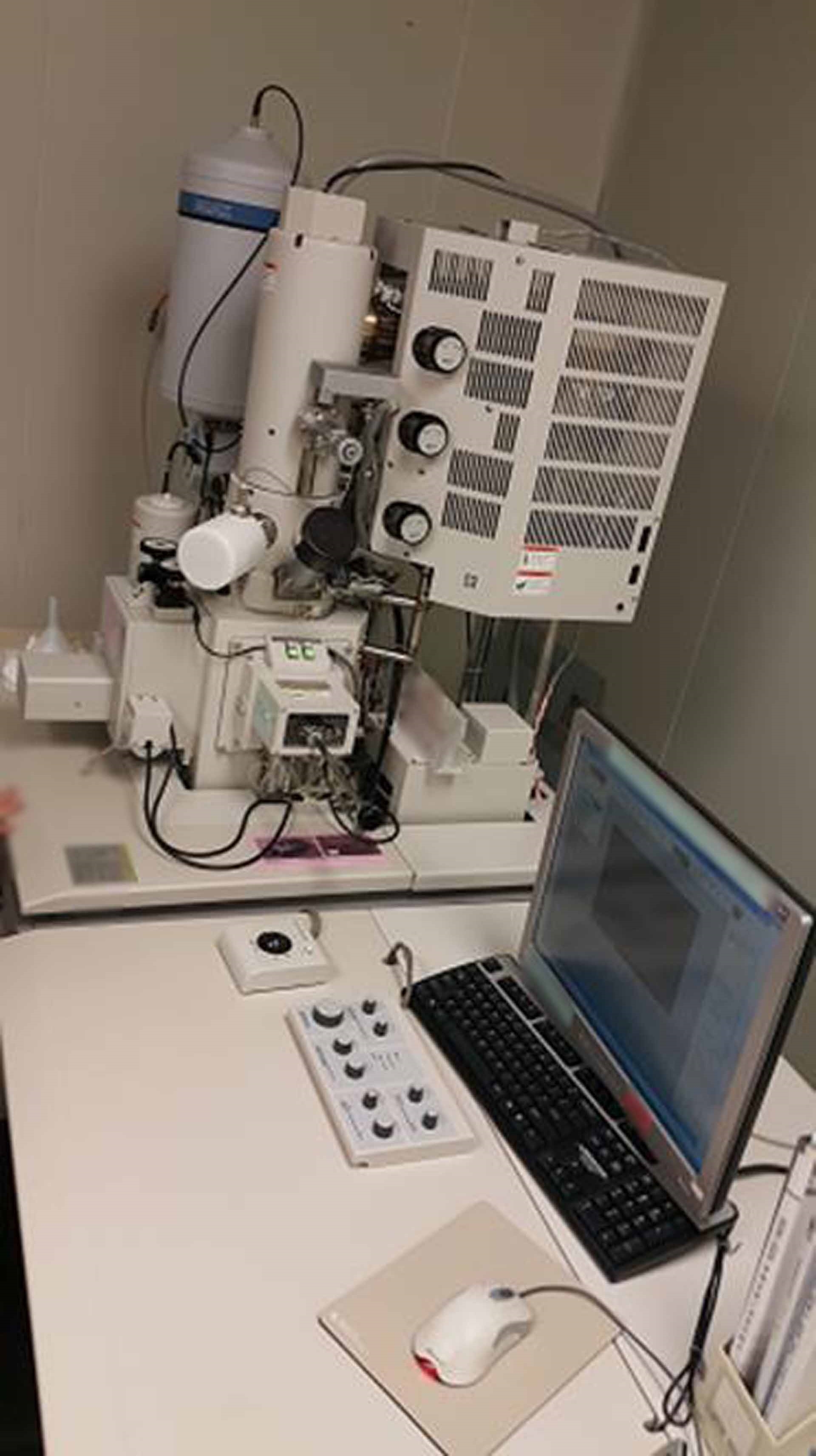
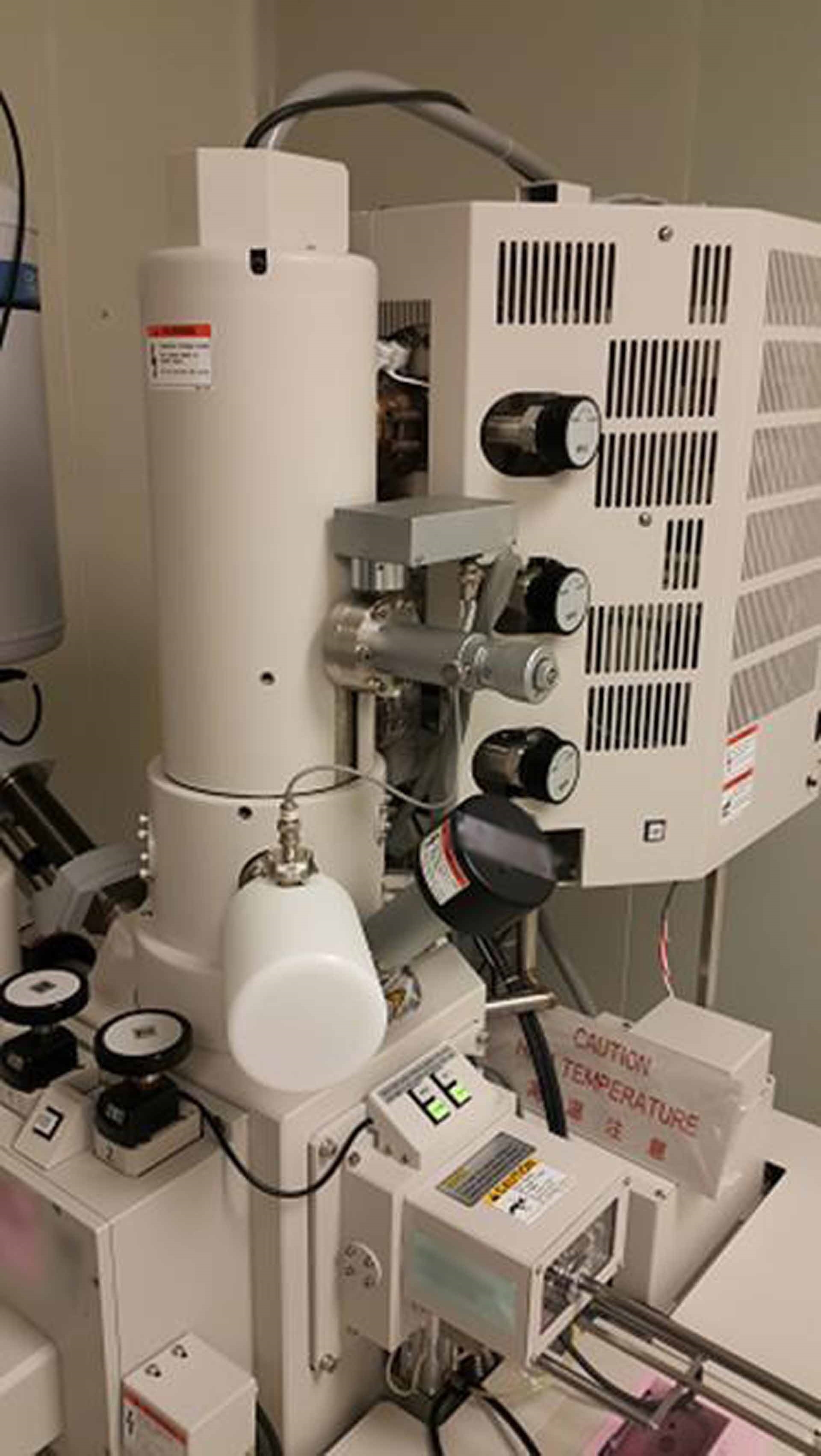
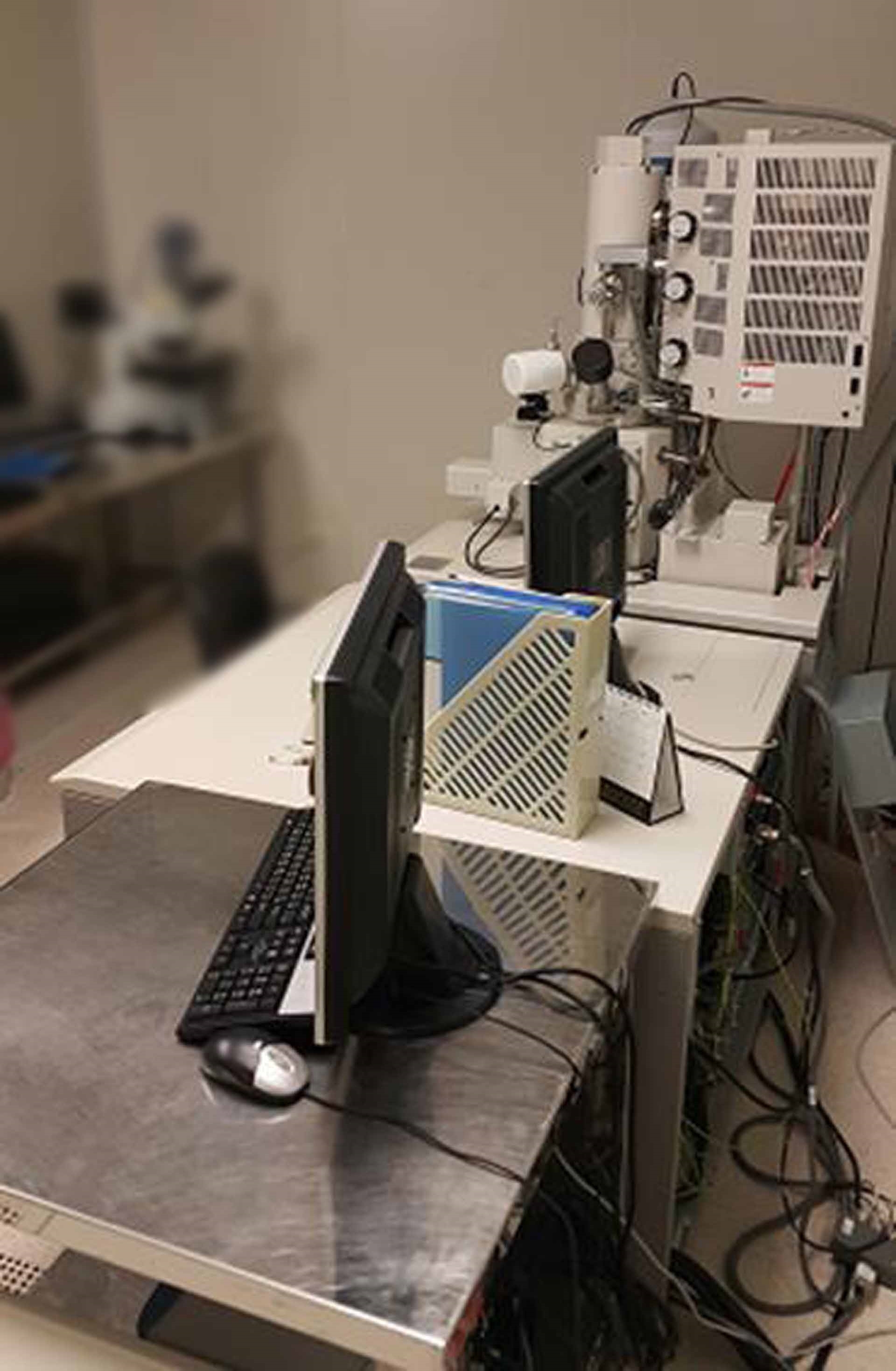
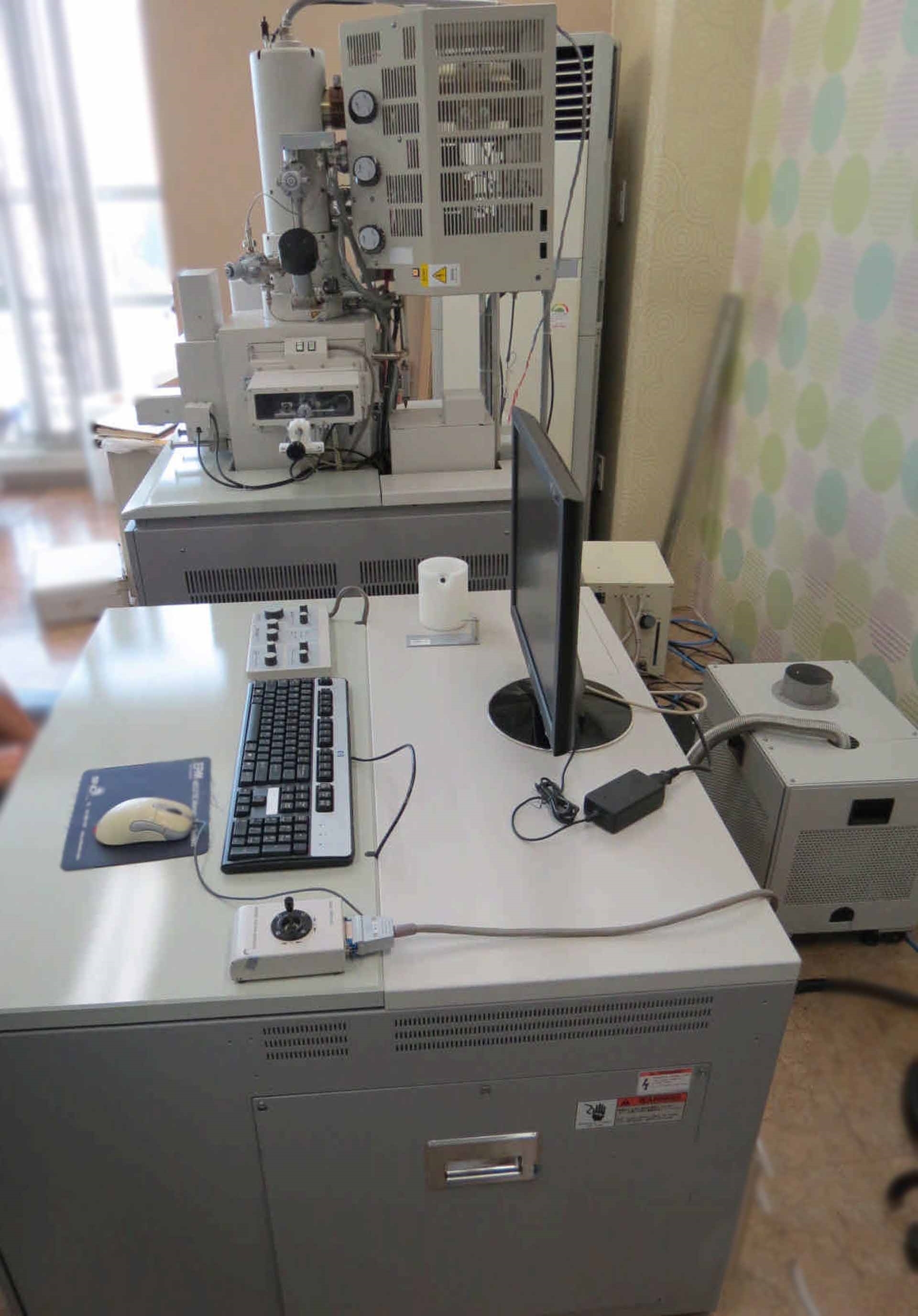
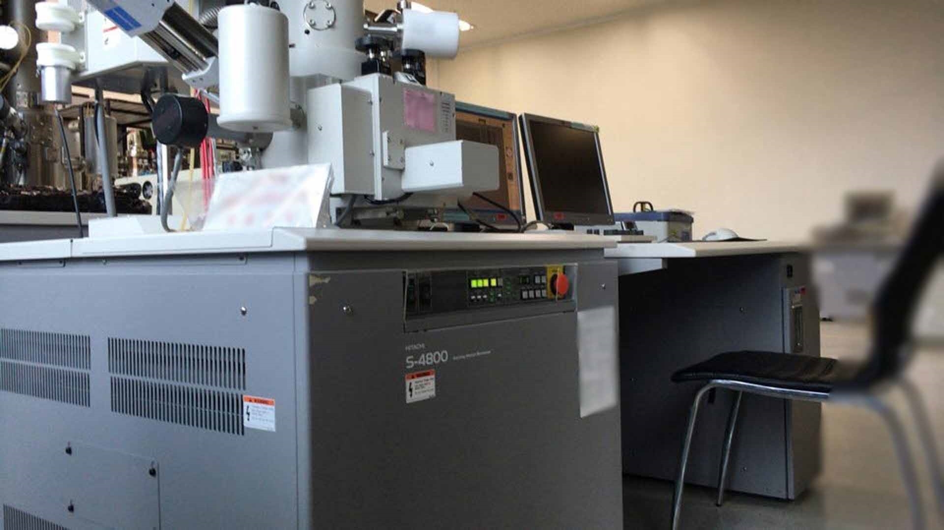
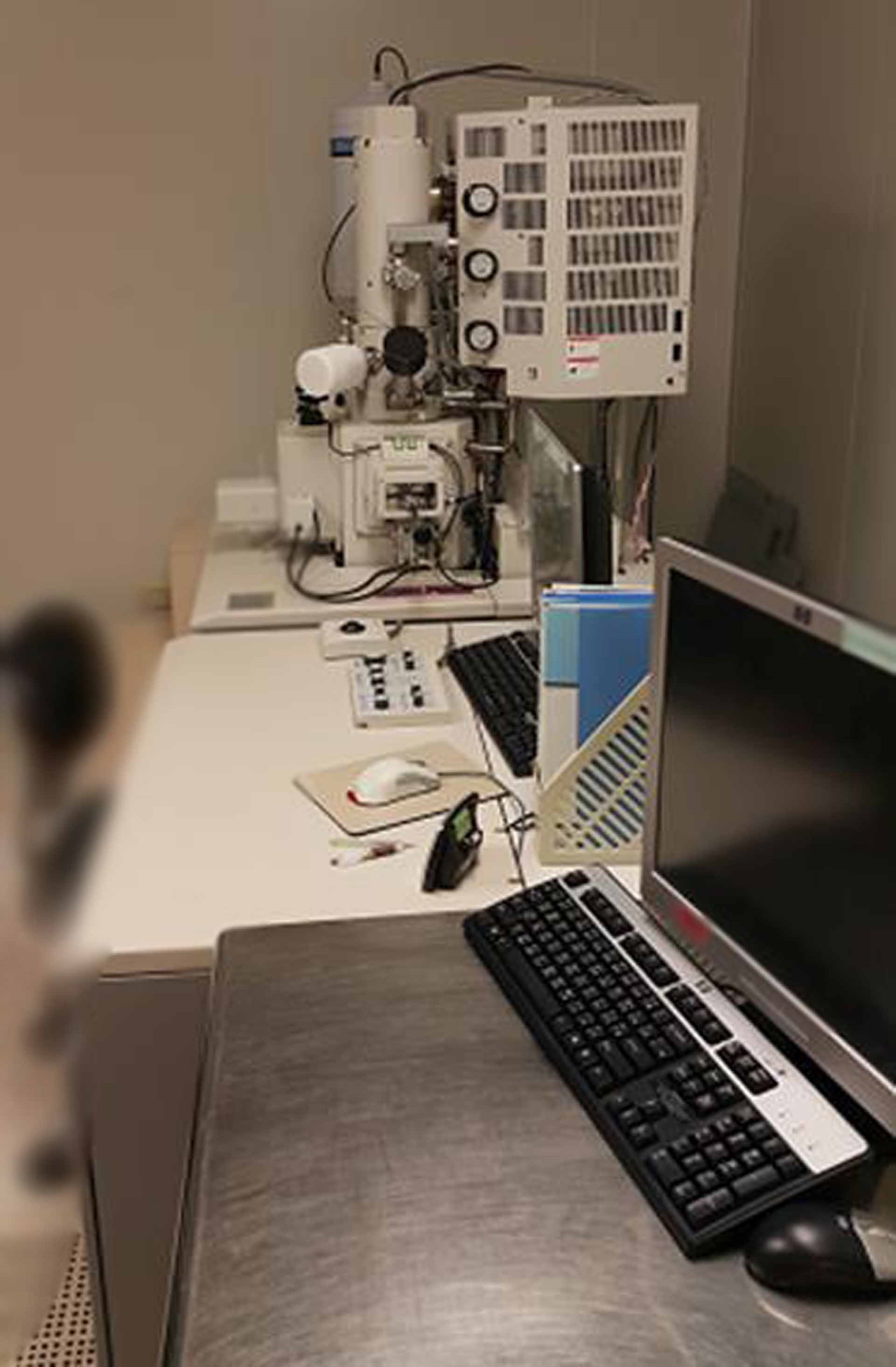
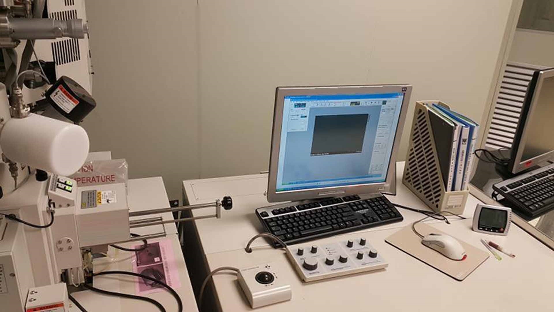
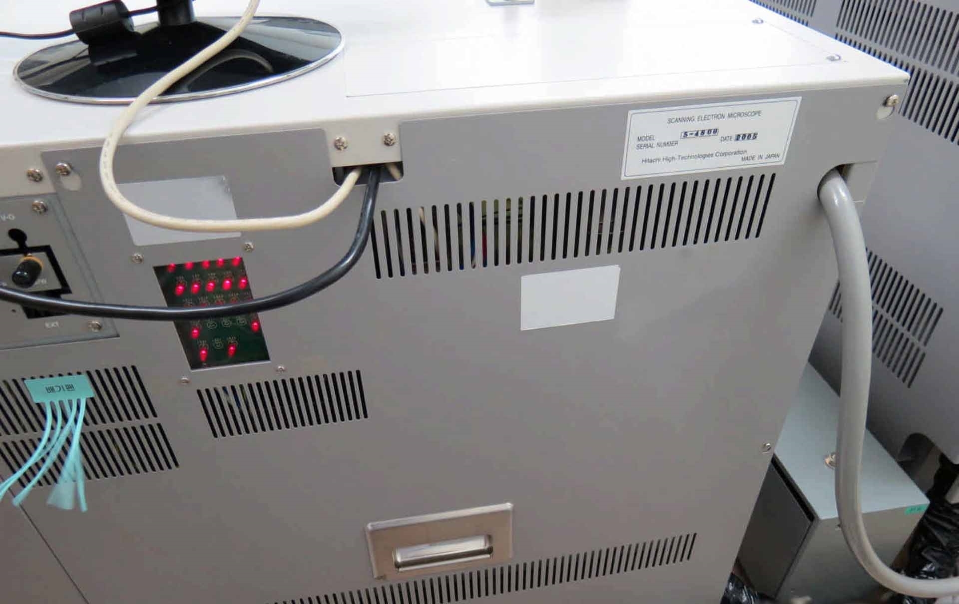
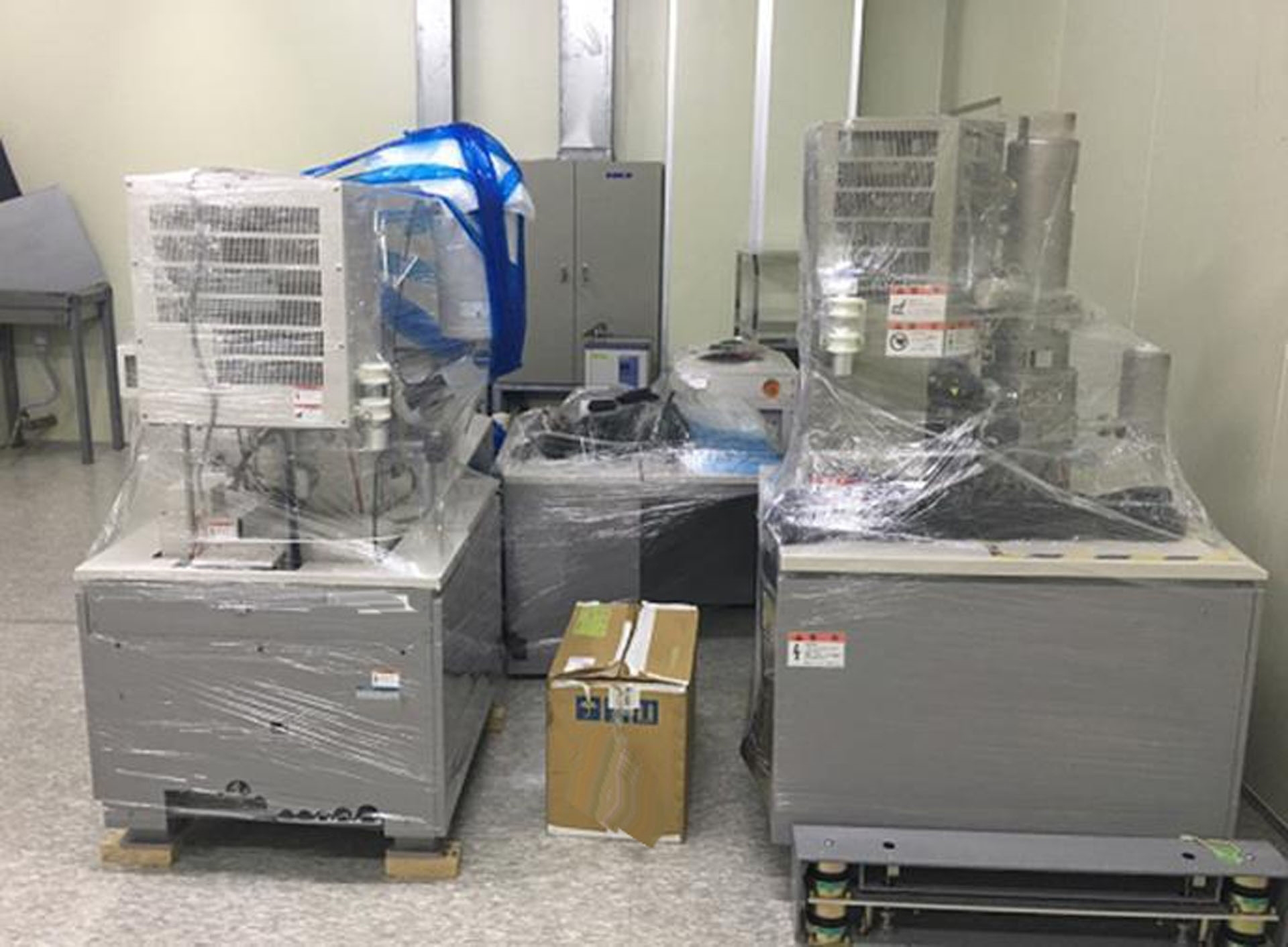
Field Emission Scanning Electron Microscope (FE-SEM)
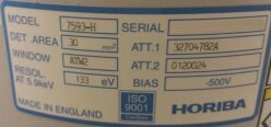
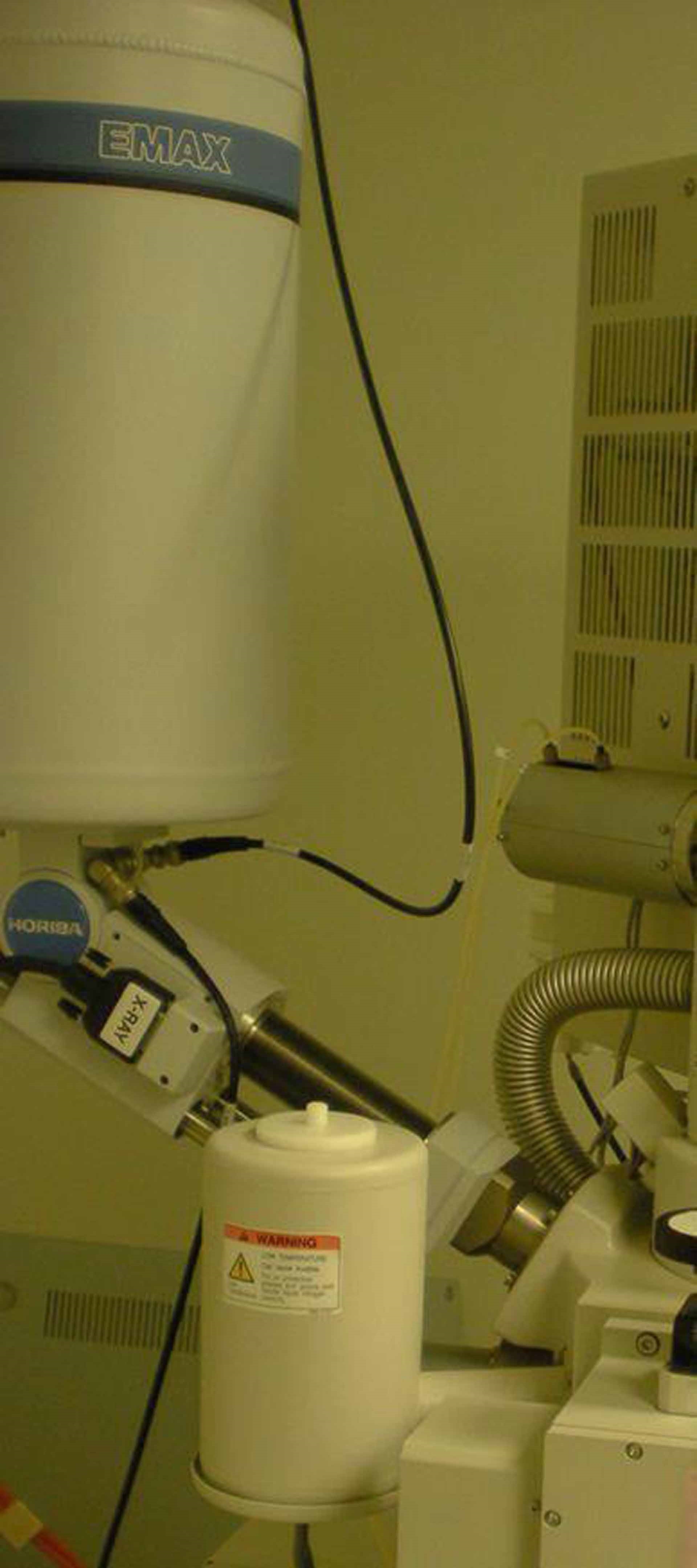
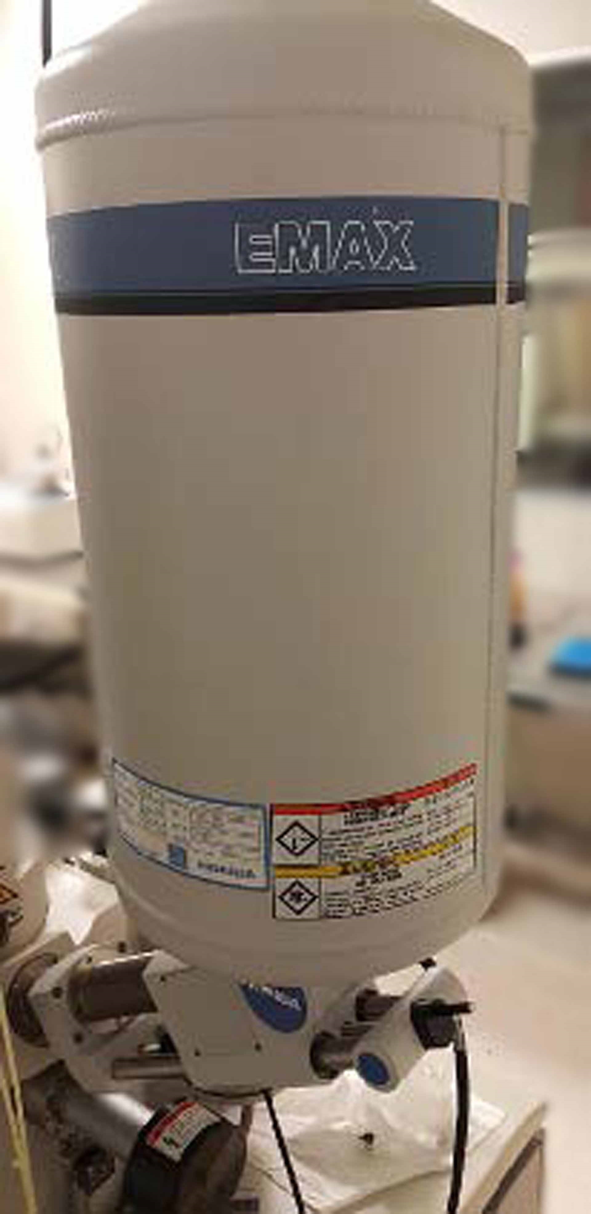
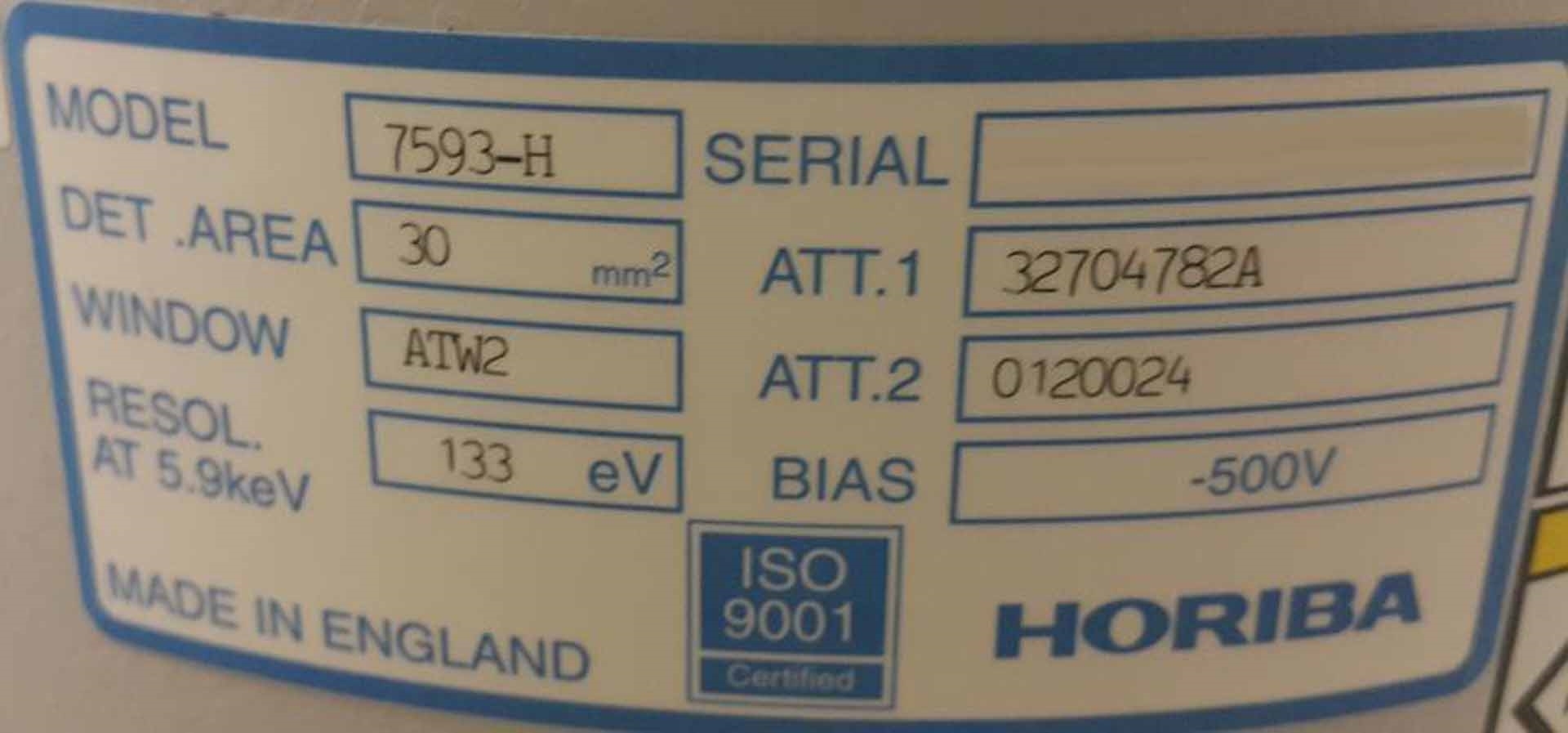
With HORIBA EDX system
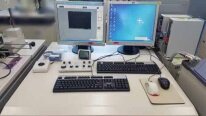
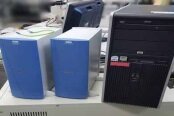
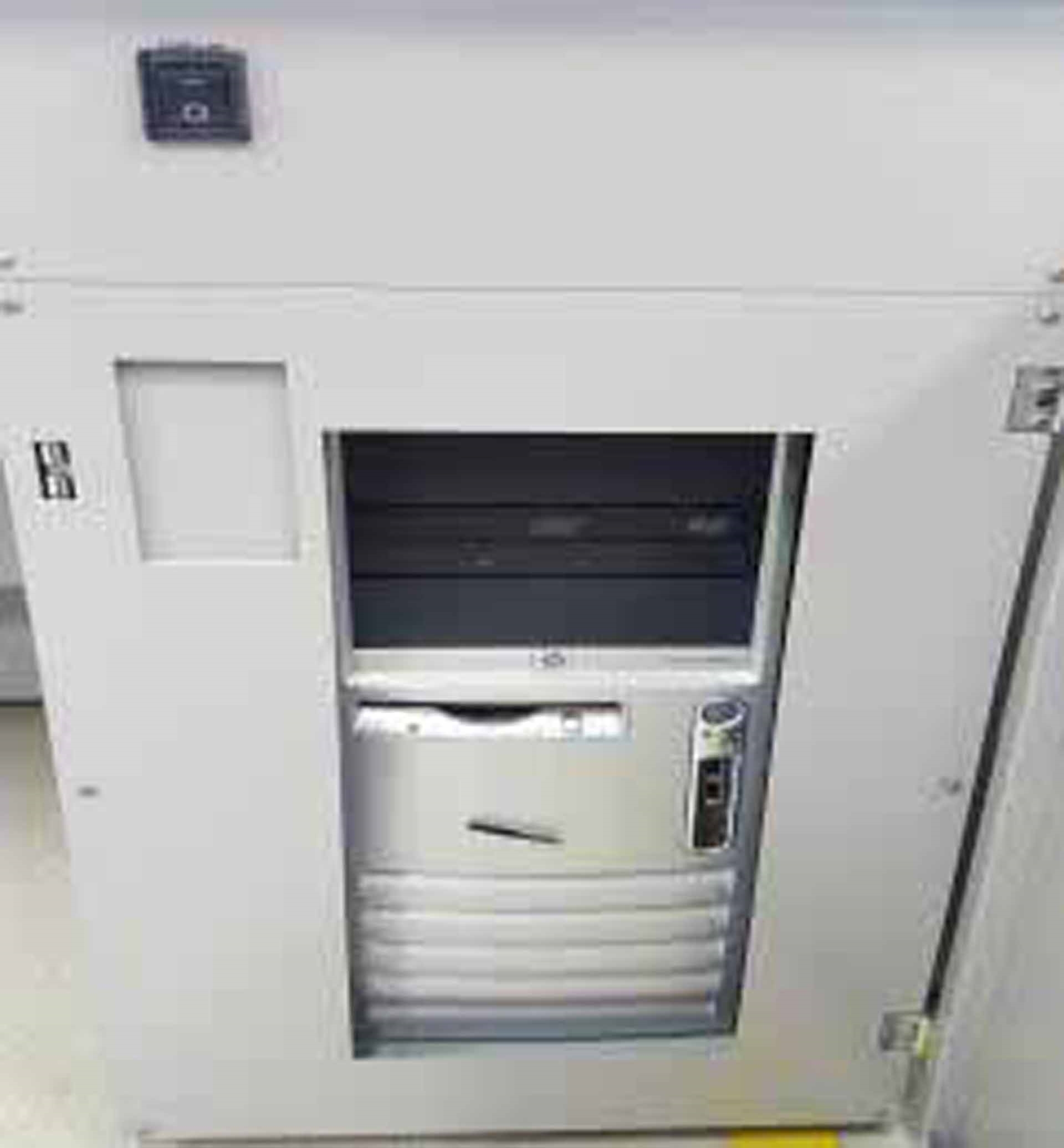
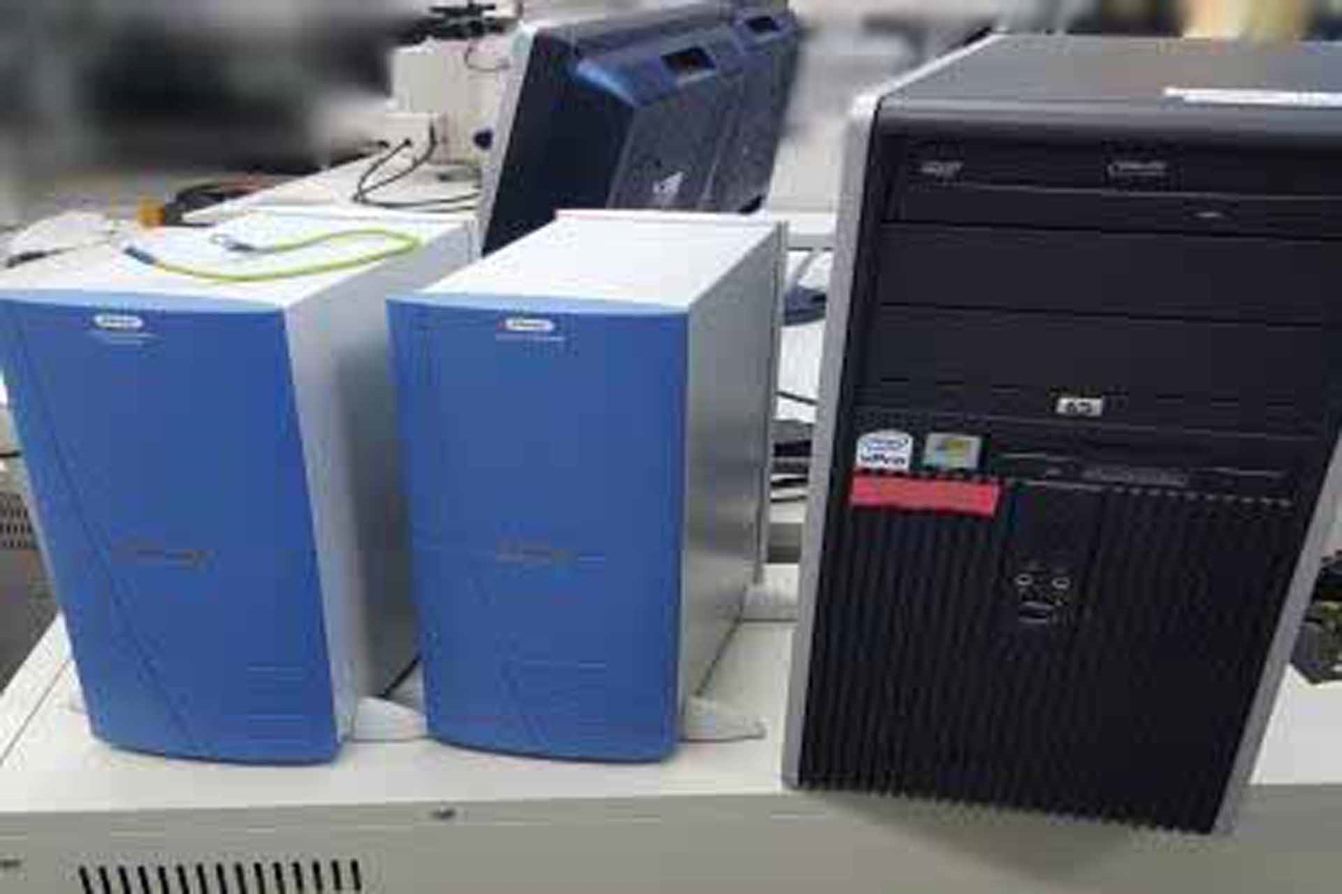
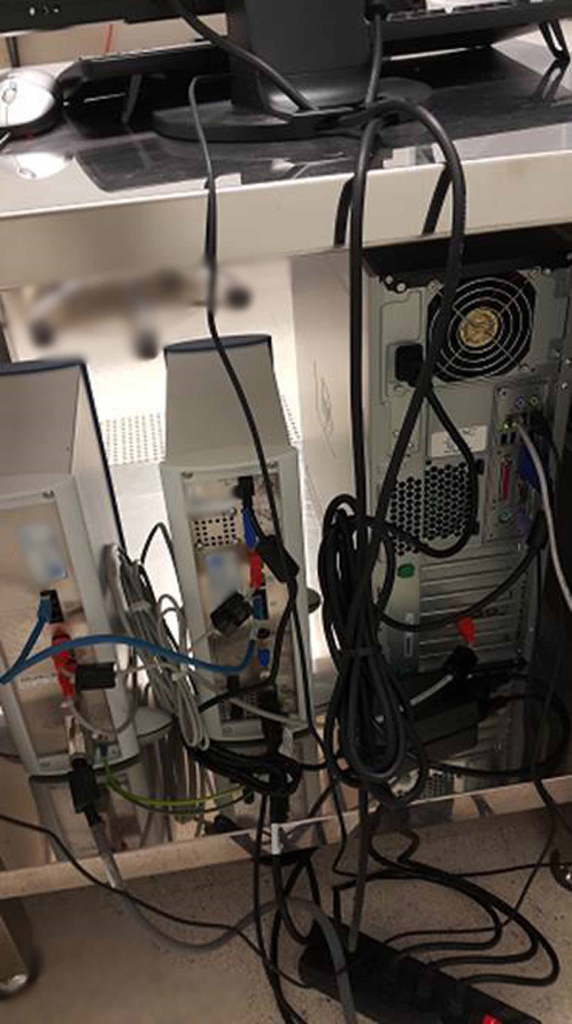
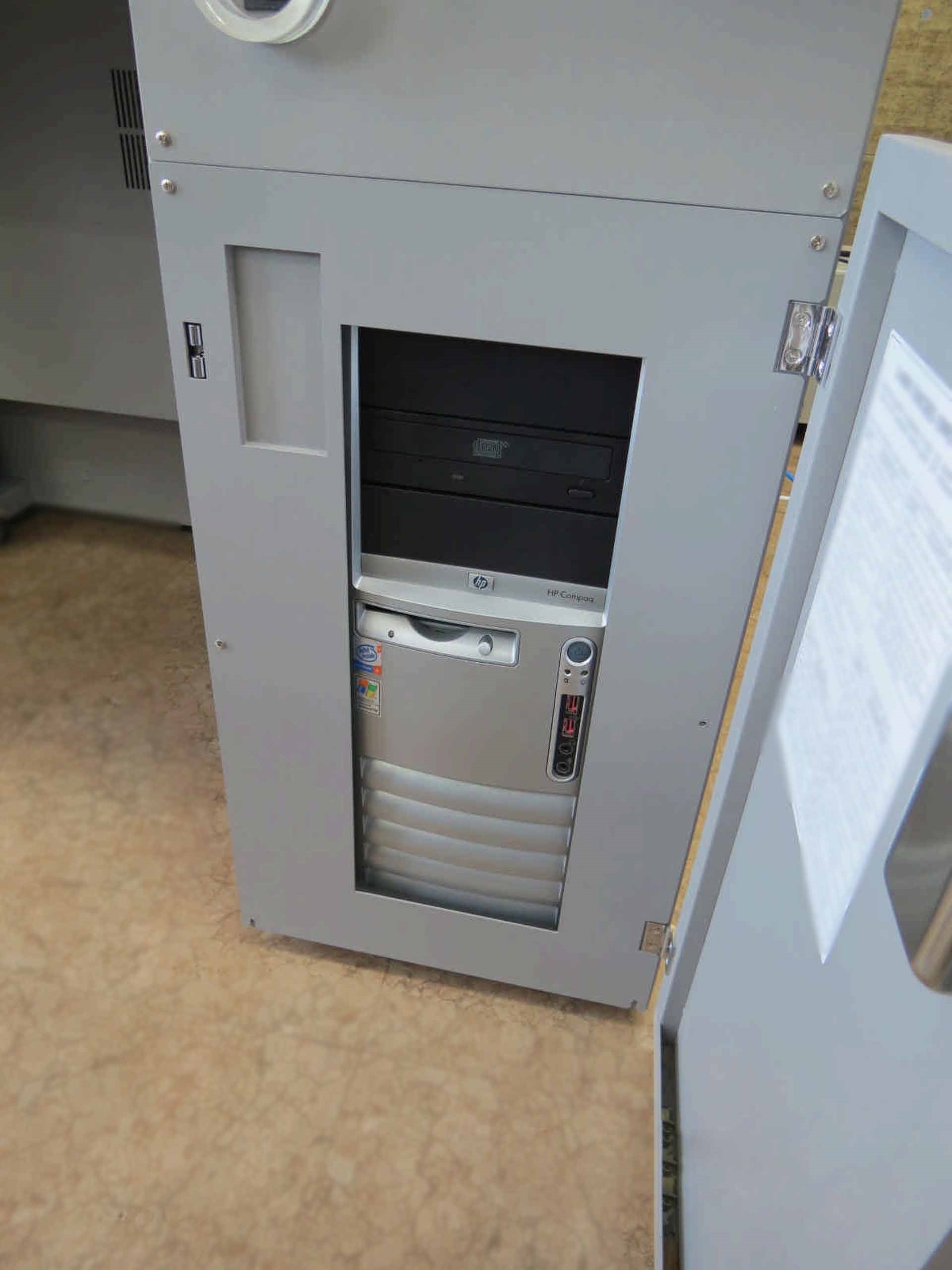
Workstation: HP DC7100MT
Declaration mode: 2.0 nm at 1 kV, WD: 1.5 mm, Normal mode
Stigmator: Octopole electromagnetic
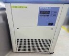
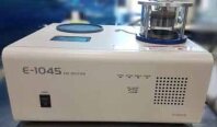
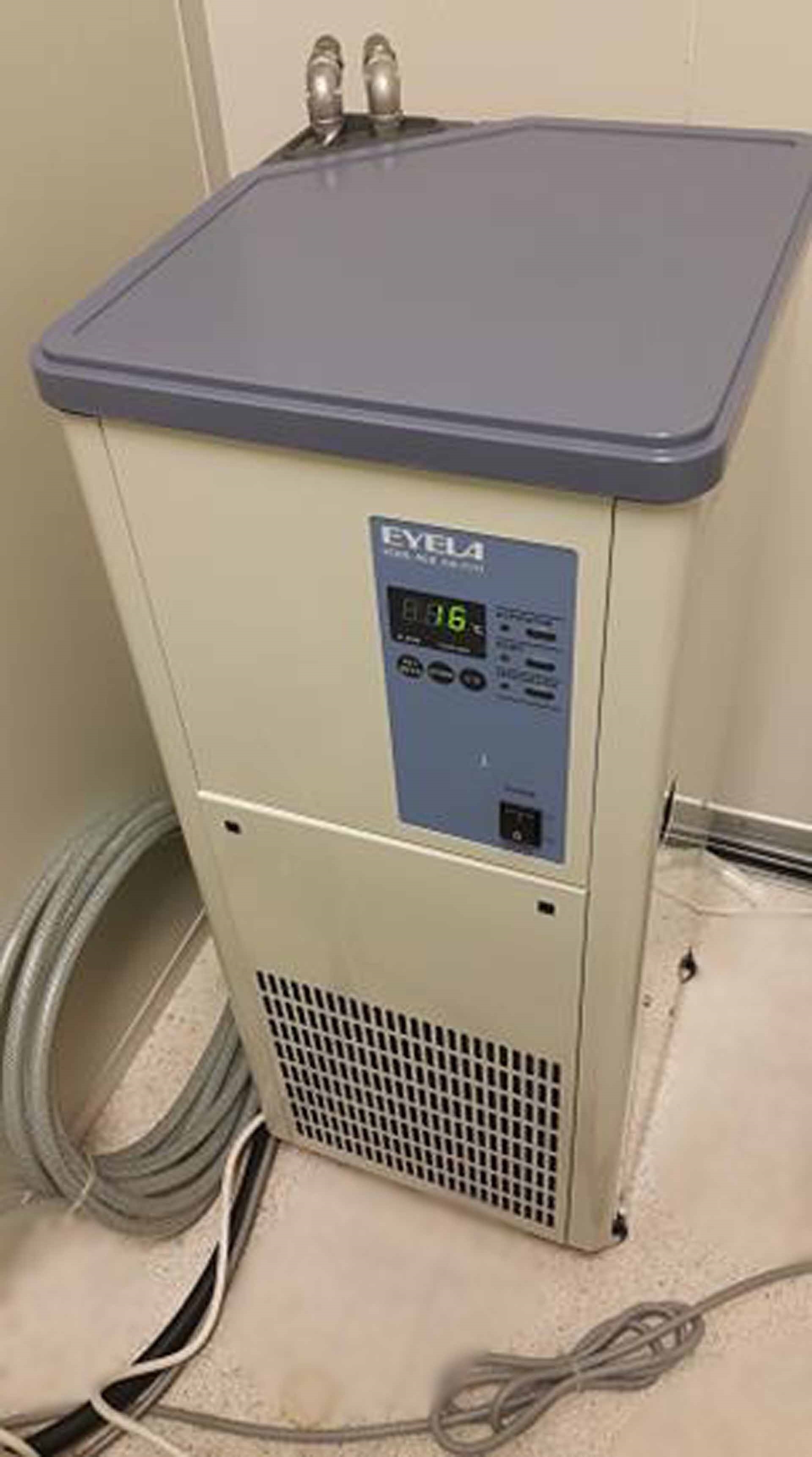
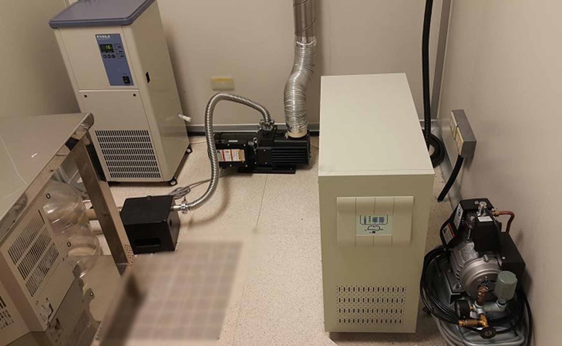
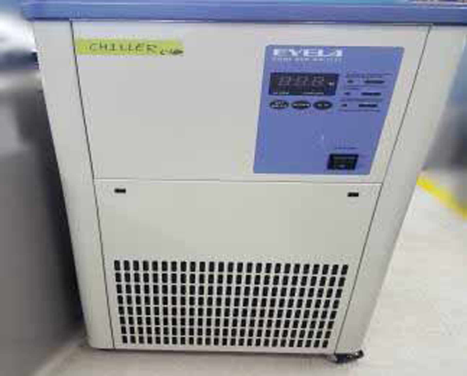
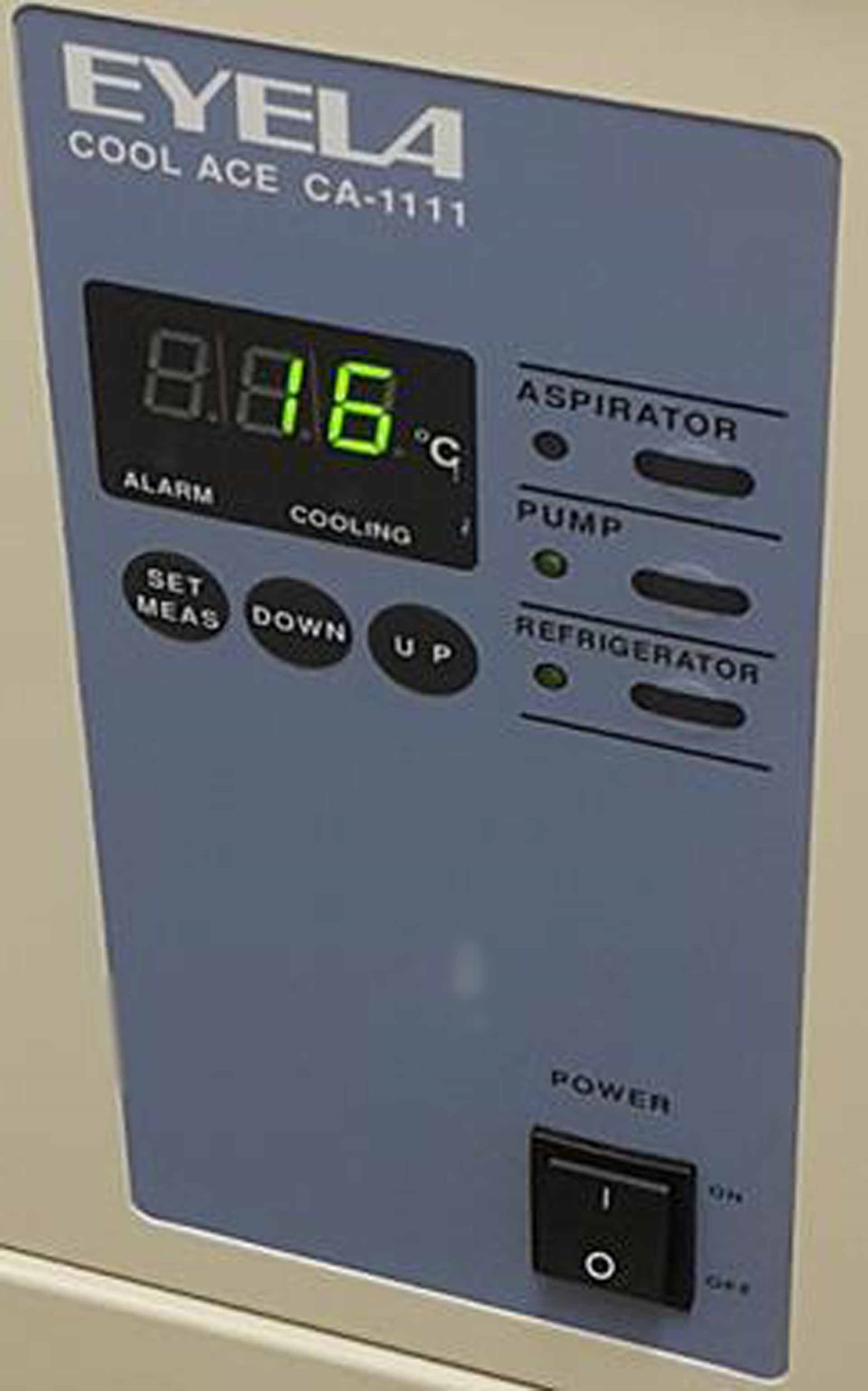
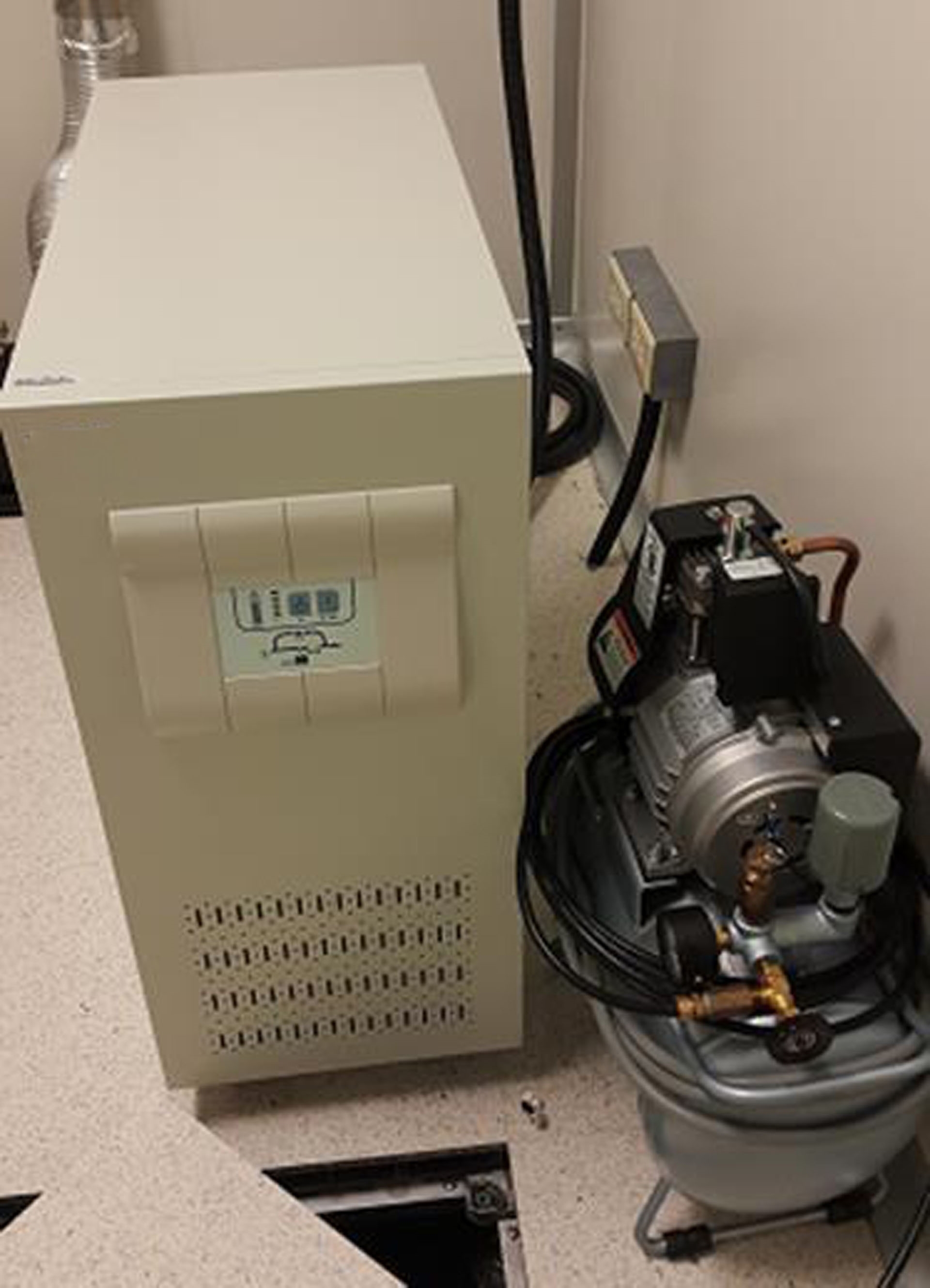
EYELA Cool Ace CA-1111 Chiller
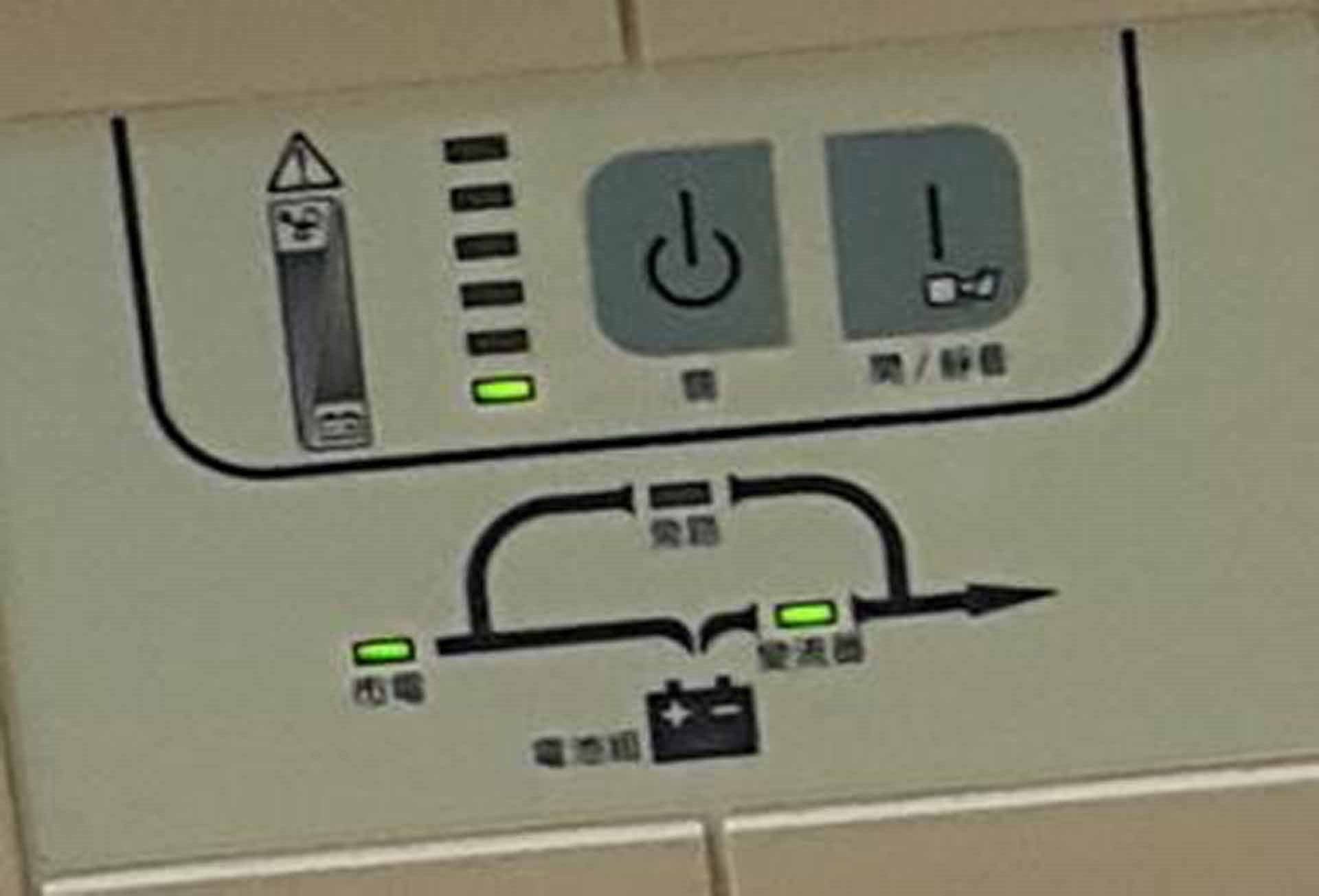
UPS
HITACHI Air compressor
Operating system: Windows XP
Resolution:
Accelerating voltage: 15 kV
Working distance: 4 mm to 1.0 nm (220,000x)
Accelerating voltage: 1 kV
Working distance: 1.5 mm to 2.0 nm (120,000x)
Magnification:
High magnification mode: 100x to 800,000x
Low magnification mode: 20x to 2,000x
Electron optics:
Electron gun: Cold cathode field emission type
Extracting voltage (Vext): 0 to 6.5 kV
Accelerating voltage (Vacc): 0.5 to 30 kV (in 100 V steps)
Lens: 3-Stage electromagnetic lens, reduction type
Objective lens aperture:
Movable aperture (4 Openings selectable / Alignable outside column)
Self-cleaning thin aperture
Astigmatism correction coil: Electromagnetic type (Stigmator)
Scanning coil: 2-Stage electromagnetic electron optics
Specimen stage:
X-Traverse: 0 to 50 mm (Continuous)
Y-Traverse: 0 to 50 mm (Continuous)
Z-Traverse: 1.5 to 30.0 mm (Continuous)
Tilt: -5° to +70°
Rotation: 360° (Continuous)
Specimen size: Max 100 mm (Diameter)
(Airlock type specimen exchange)
Display unit:
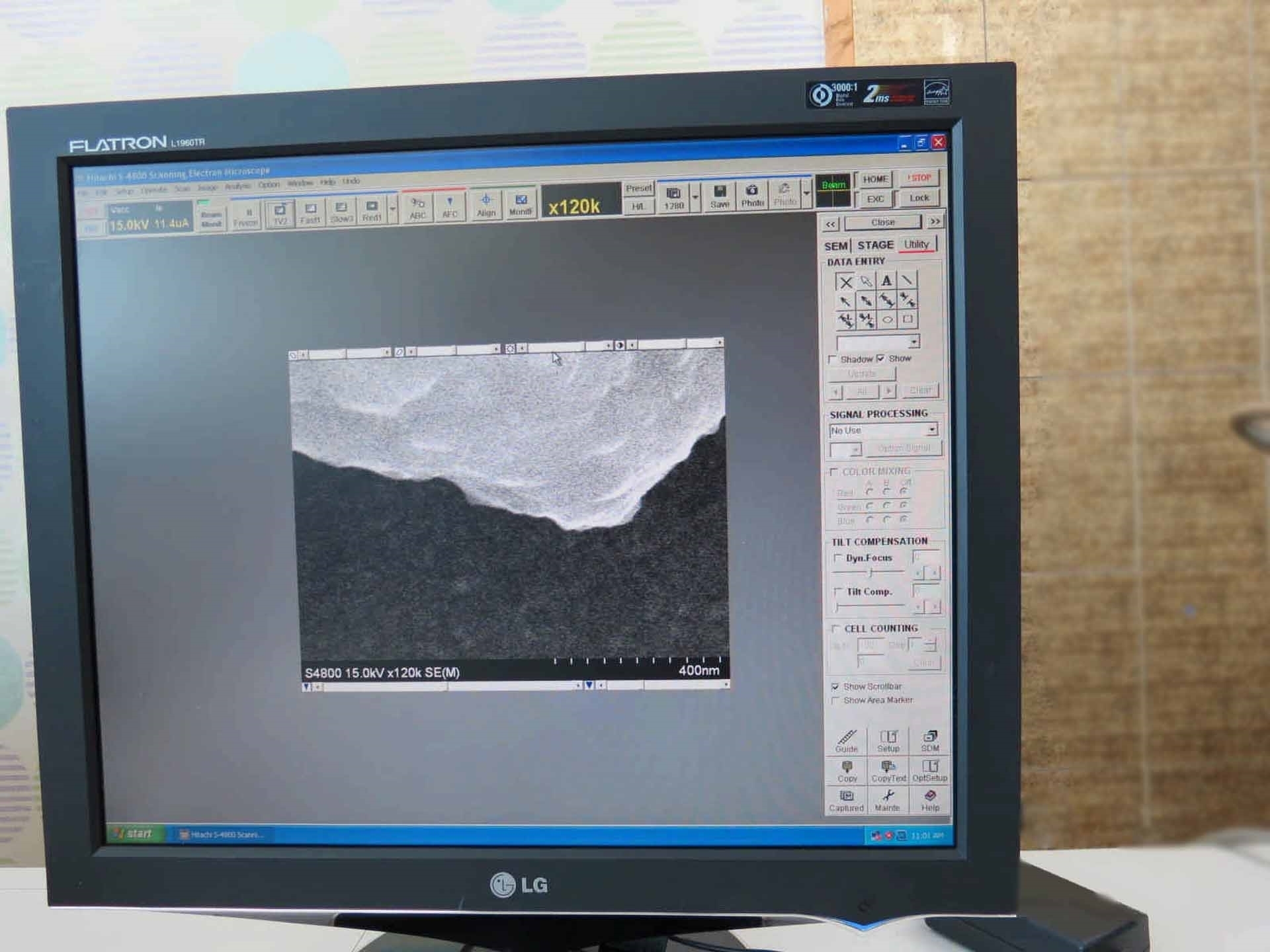
Display type: Flicker-free image on PC monitor (Full scanning speeds)
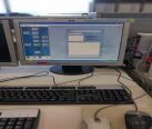
Viewing monitor: Type 18.1 LCD
Option: Type 21 Color CRT (1280 x 1024 pixels)
Photo CRT (Option): Ultra-high resolution type
(Effective field of view 120 x 90 mm)
Full screen: 1280 x 960 Pixels
Reduced area:
640 x 480 Pixels
320 x 240 Pixels
Dual Image: 640 x 480 Pixels
Scanning modes:
Normal scan
Reduced area scan
Line scan
Spot analysis
Average concentration analysis
Split / Dual magnification
Scanning speeds:
TV (640 x 480 pixel display: 25 / 30 frames/s)
Fast (Full screen display: 6.25 / 7.5 frames/s)
Slow:
(Full screen display: 1 / 0.9 , 4 / 3.3 , 20 / 16, 40 / 32 ,80 / 64 Frames/s)
(640 x 480 pixels display: 0.5 / 0.4 , 2 / 1.7 , 10 / 8 , 20 / 16, 40 / 32 Frames/s)
Photograph: 2560 x 1920 Pixels
Display: 40 / 32, 80 / 64, 160 / 128, 320 / 256 Frames/s
Value: 50 Hz / 60 Hz
TV: NTSC or PAL Signal
Signal processing modes:
Automatic brightness control
Gamma control
Automatic focus
Automatic stigmator
Automatic data display:
Image number
Accelerating voltage
Magnification
Micron bar
Micron value
Data / Time
Data entry: Alphanumeric characters, number, and marks
Electrical image shift: 12 m (WD: 8 mm)
Evacuation system:
System type: Fully automatic pneumatic-valve system
Ultimate vacuum levels: Specimen chamber: 7 x Pa
10-7 Pa in electron gun chamber
10-4 Pa in specimen chamber
Electron gun chamber:
IP1 1 x Pa or better
IP2 2 x Pa or better
IP3 7 x Pa or better
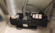
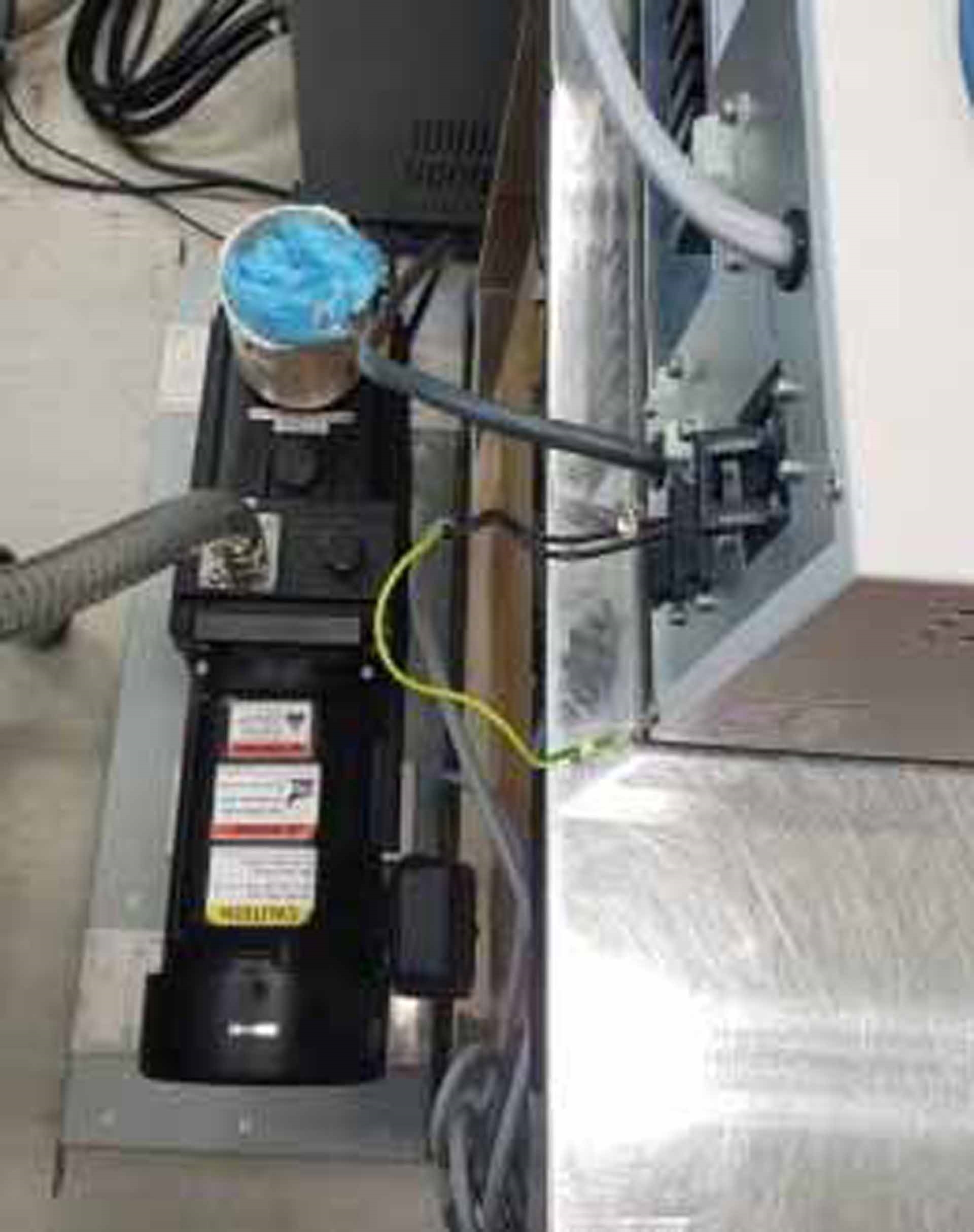
Vacuum pumps: ULVAC GLD-136
Electron optical system: (3) Ion pumps
Specimen chamber: Turbo molecular pump
Oil rotary pump
Protection devices:
Warning devices:
Power failure
Cooling-water interruption
Inadequate vacuum
Secondary electron image resolution:
1.0 nm at 15 kV, WD: 4 mm
1.4 nm at 1 kV, WD: 1.5 mm
Sample chamber:
Size: Type I stage
Max sample size: 100 mm Diameter
Stage motion:
3 Axis motorized
X/Y: 0 - 50 mm
Signal selection:
SE Signal
X-Ray signal
AUX Signal
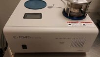
UPS Unit
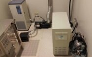
Chiller
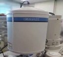
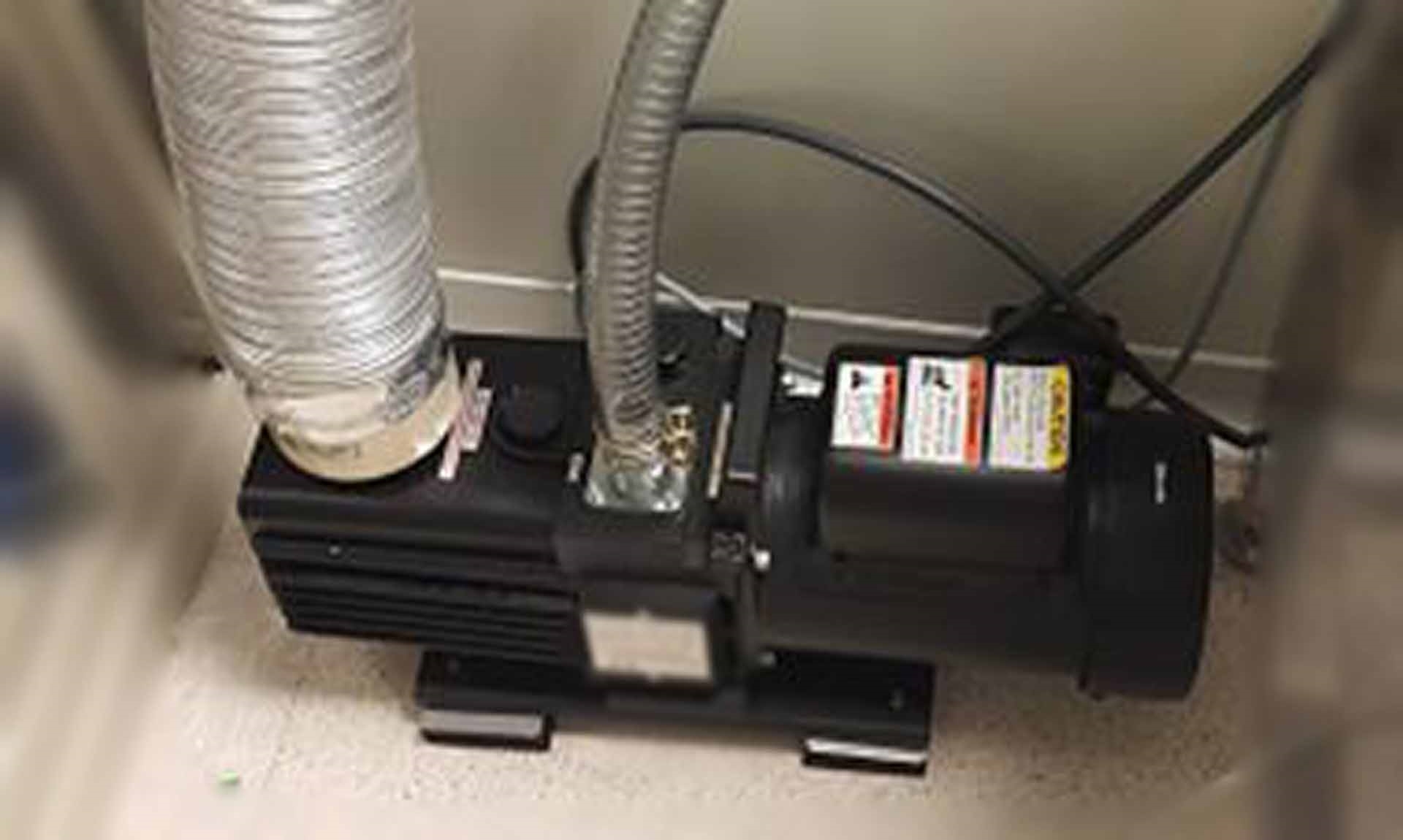
FE-SEM Rotary pump
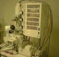
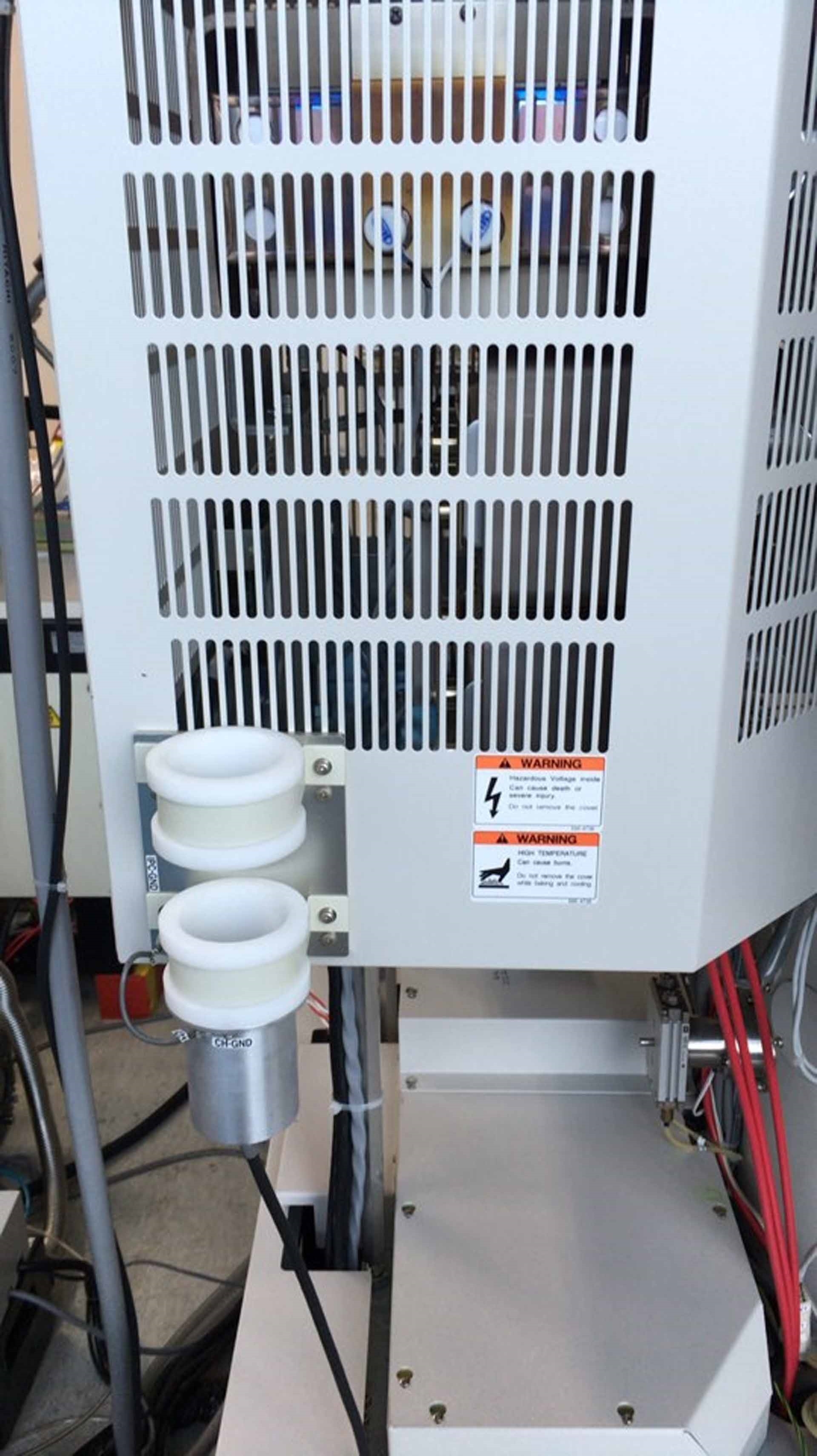
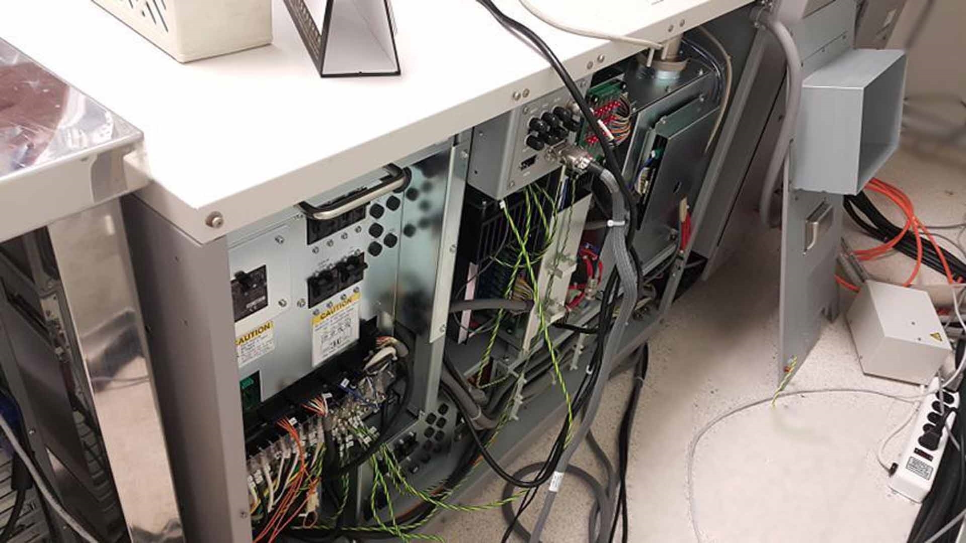
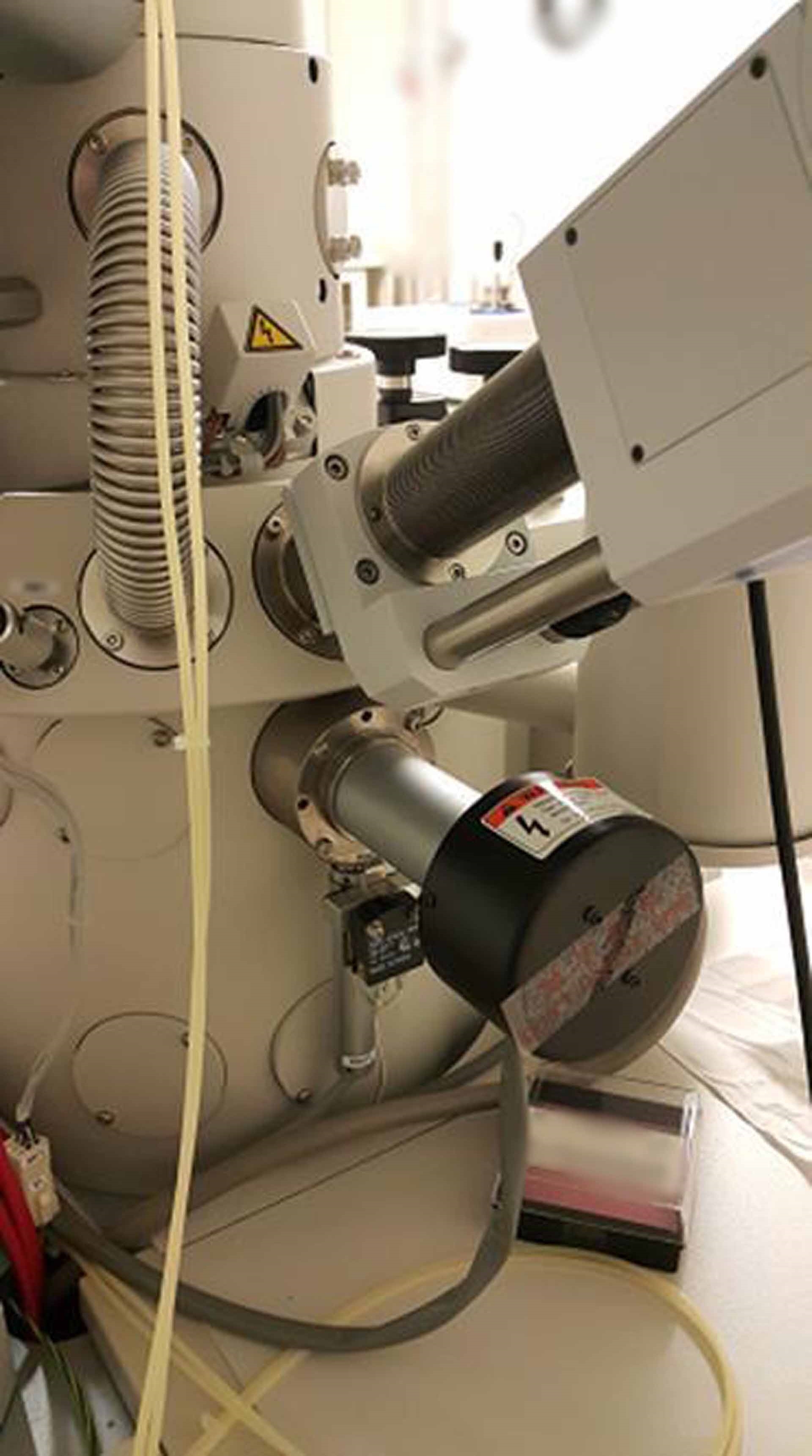
Main body:
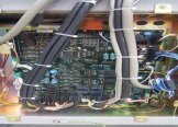
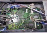
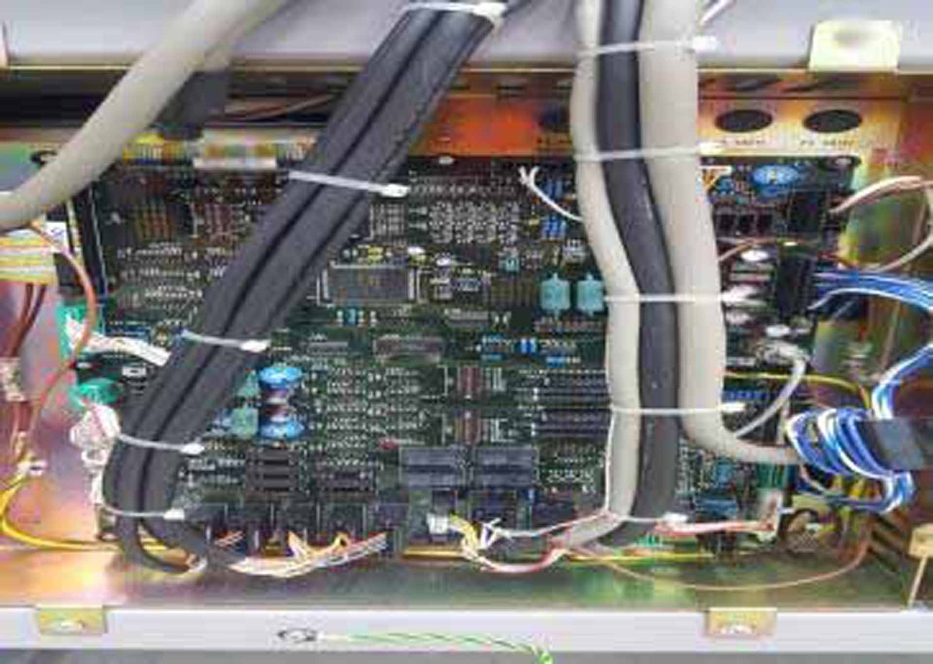
Stage controller (ROM PCB)
EVAC Controller (ROM PCB)
(3) Ion pumps
SE Detector
Multi-aperture
Gun head cap
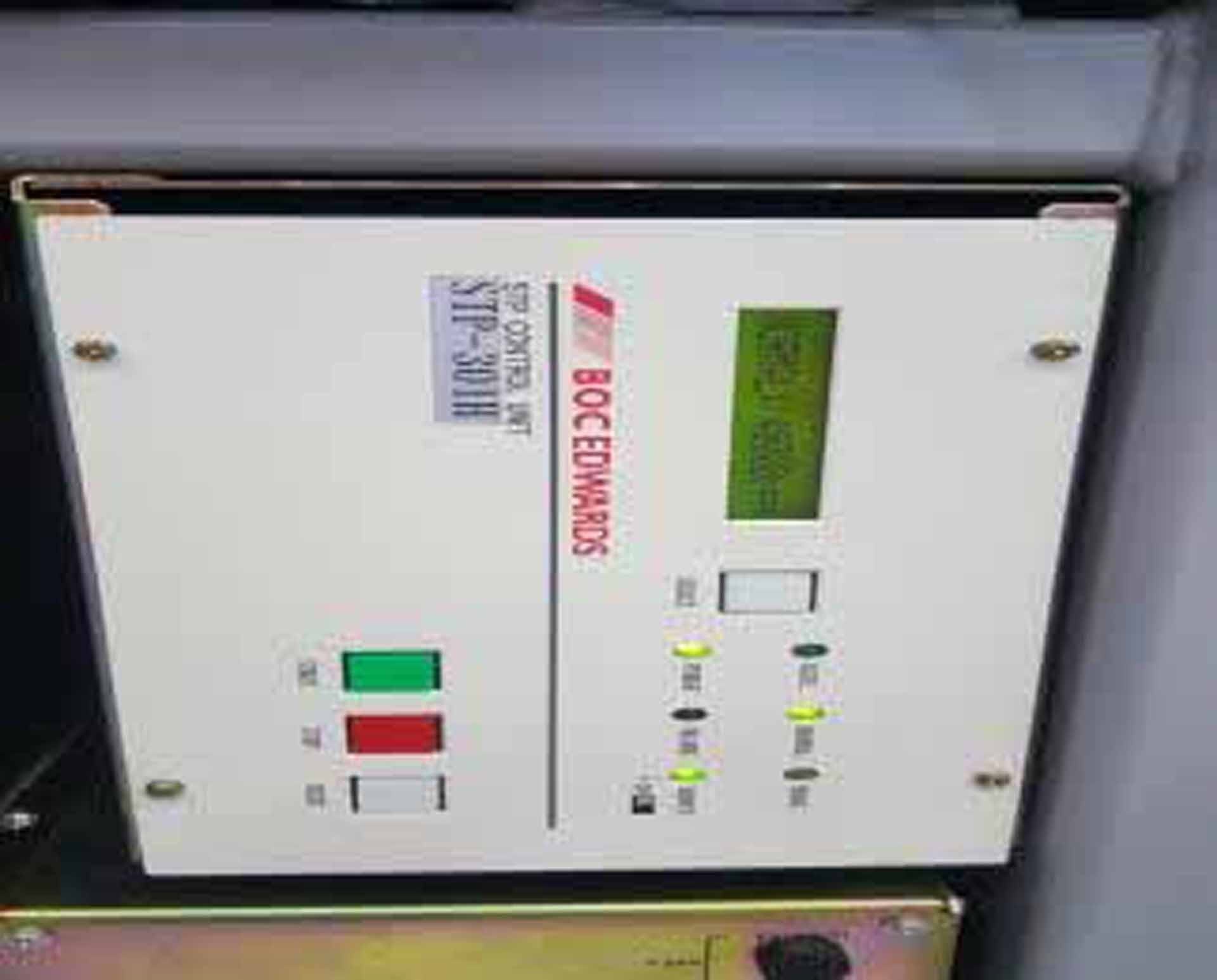
STP301H TMP Pump
STP301H TMP Controller
Solenoid valve assy
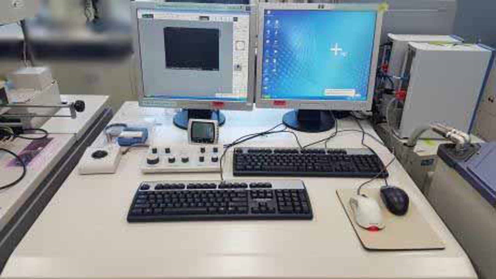
PC
Hard Disk Drive (HDD)
Operating system: Windows XP
HV Controller
Gun head unit
Case
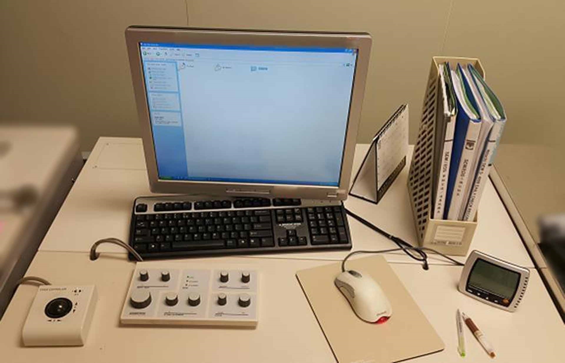
Operation unit:
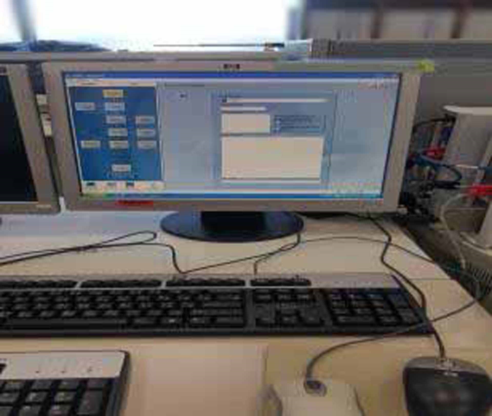
LCD Monitor
Keyboard
Mouse
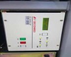
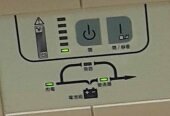
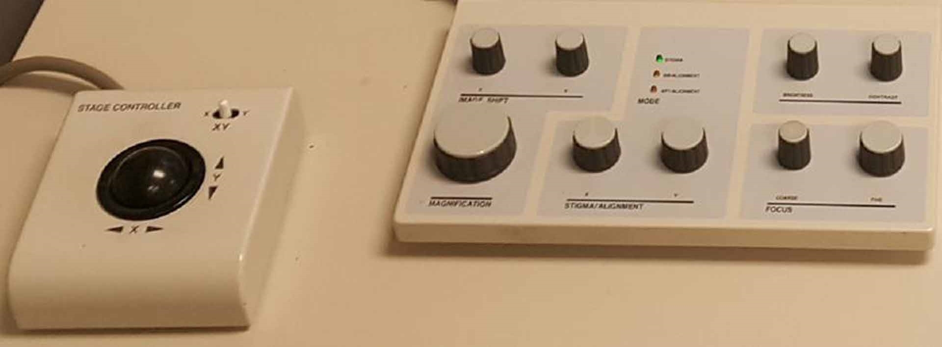
Operation panel
Stage control trackball
ETC:
Ion coater rotary pump
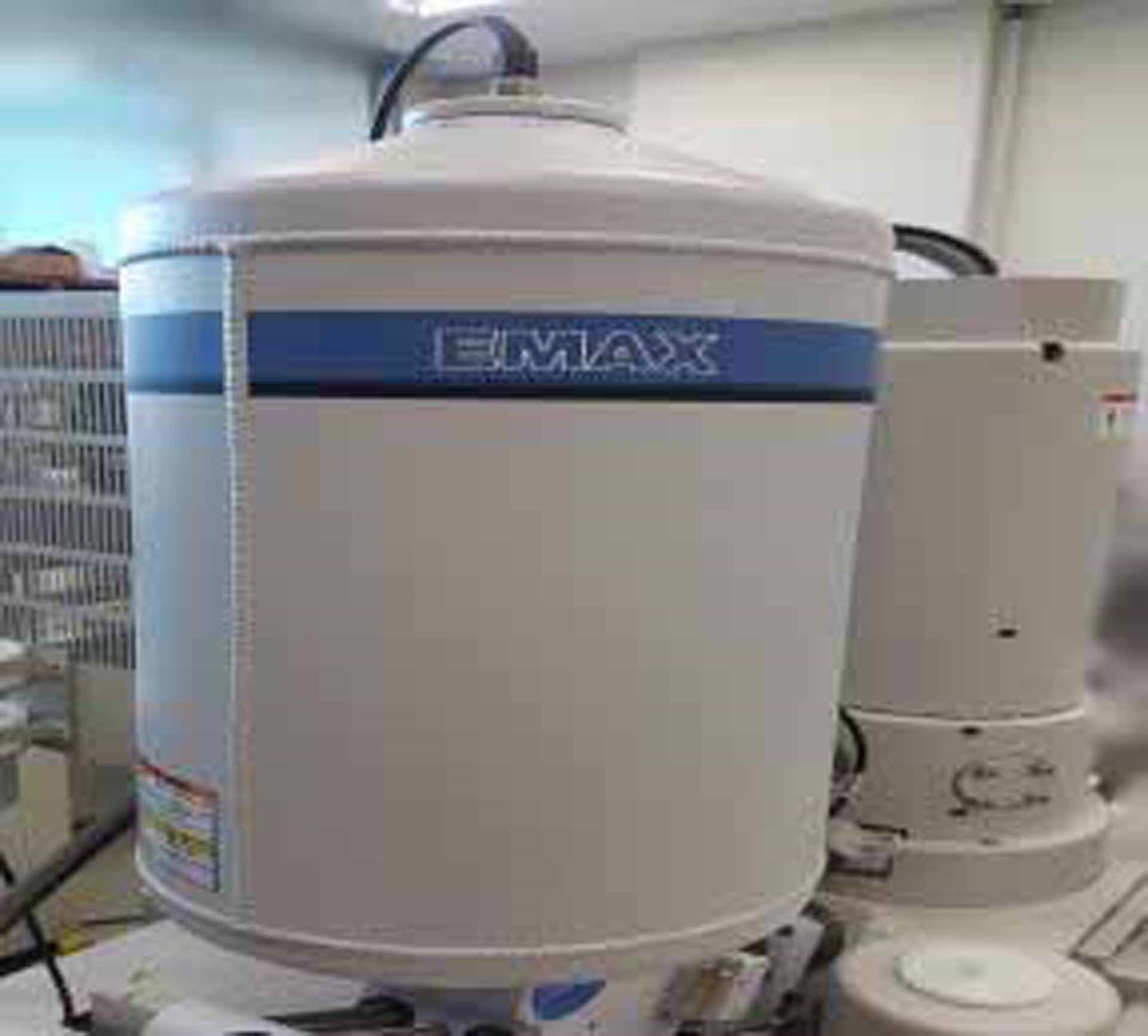
LN2
UPS
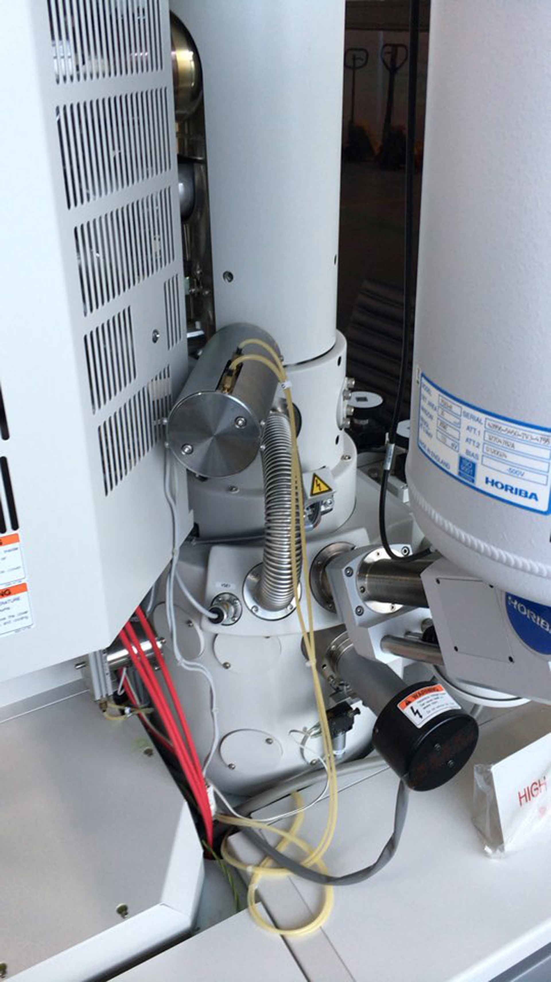
EDS
Control HUB
EDS Controller
Operating system: Windows XP
Baking tool
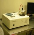
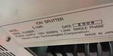
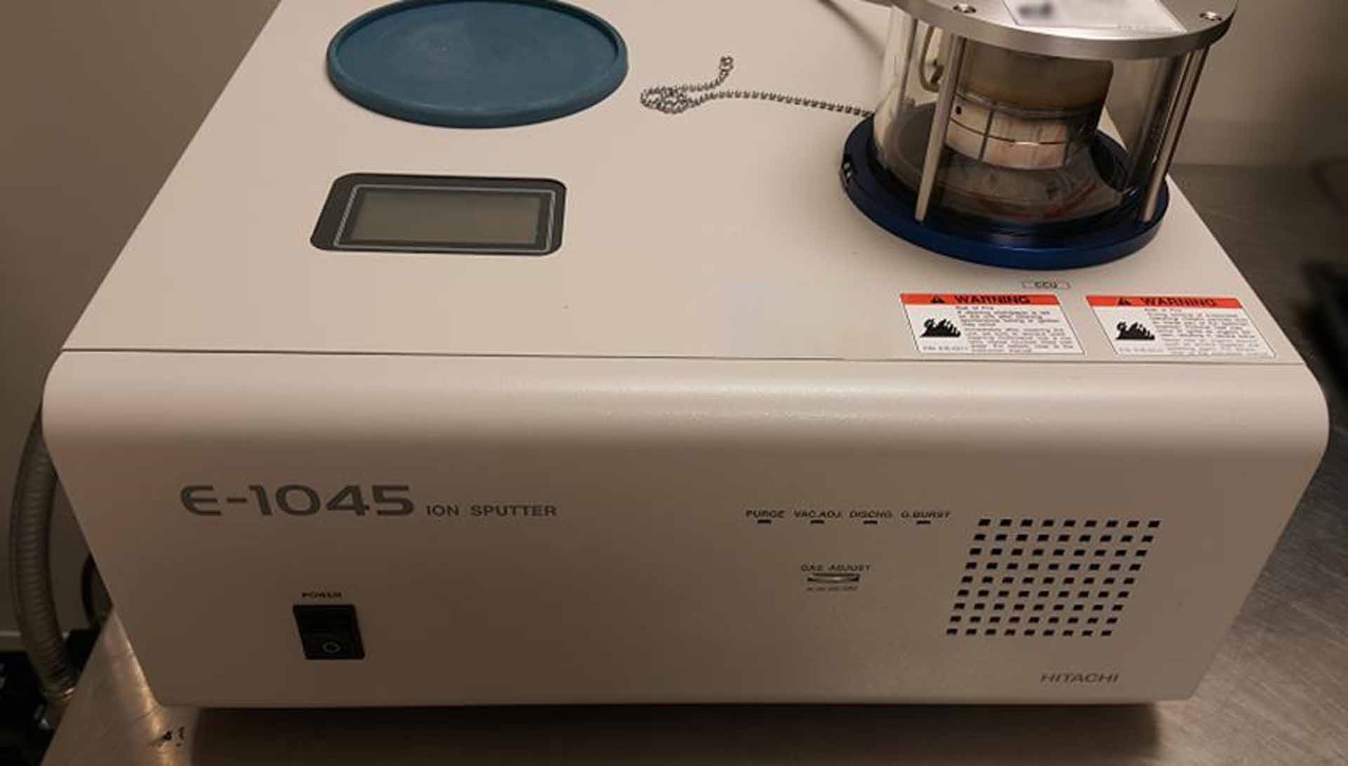
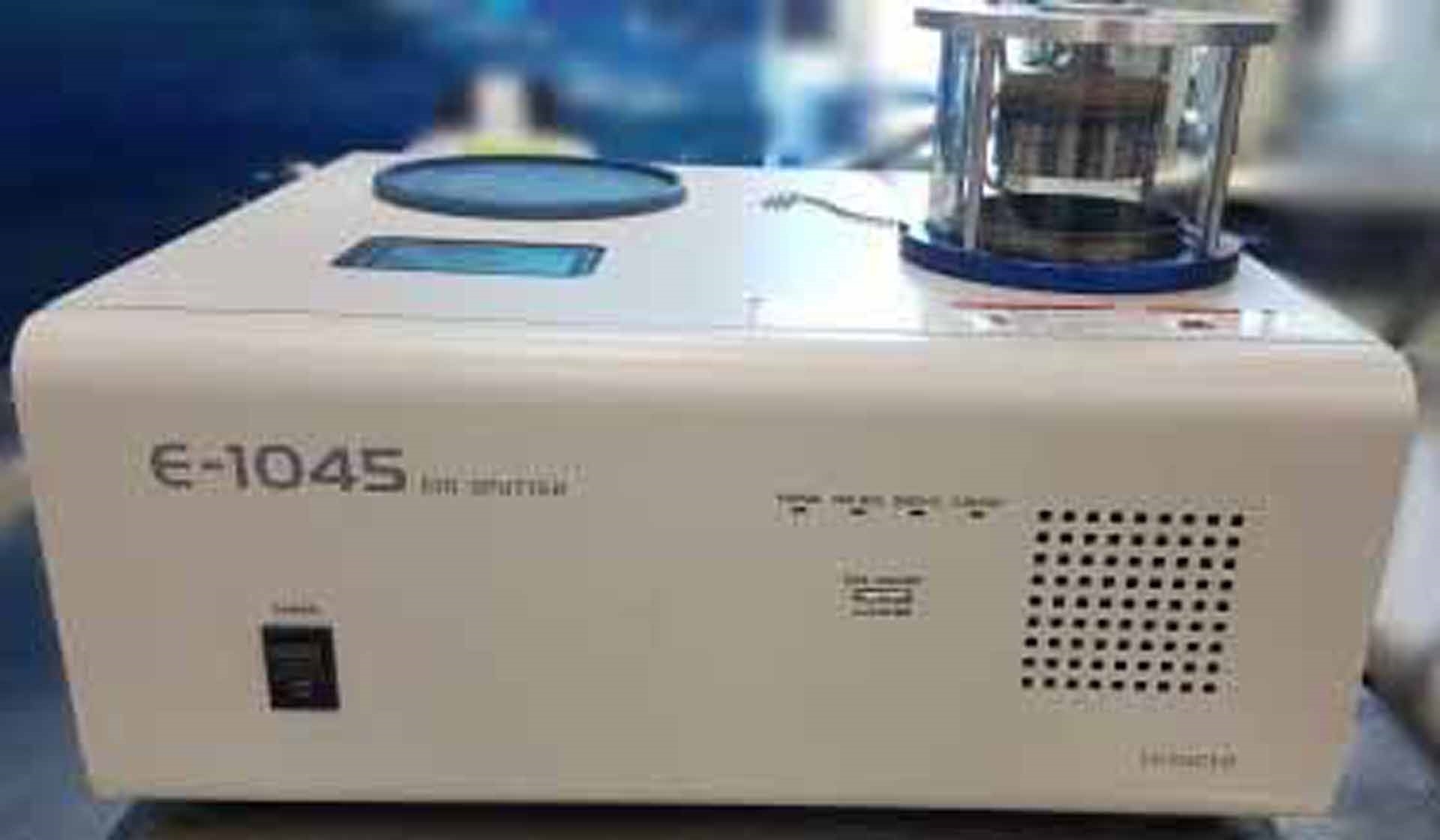
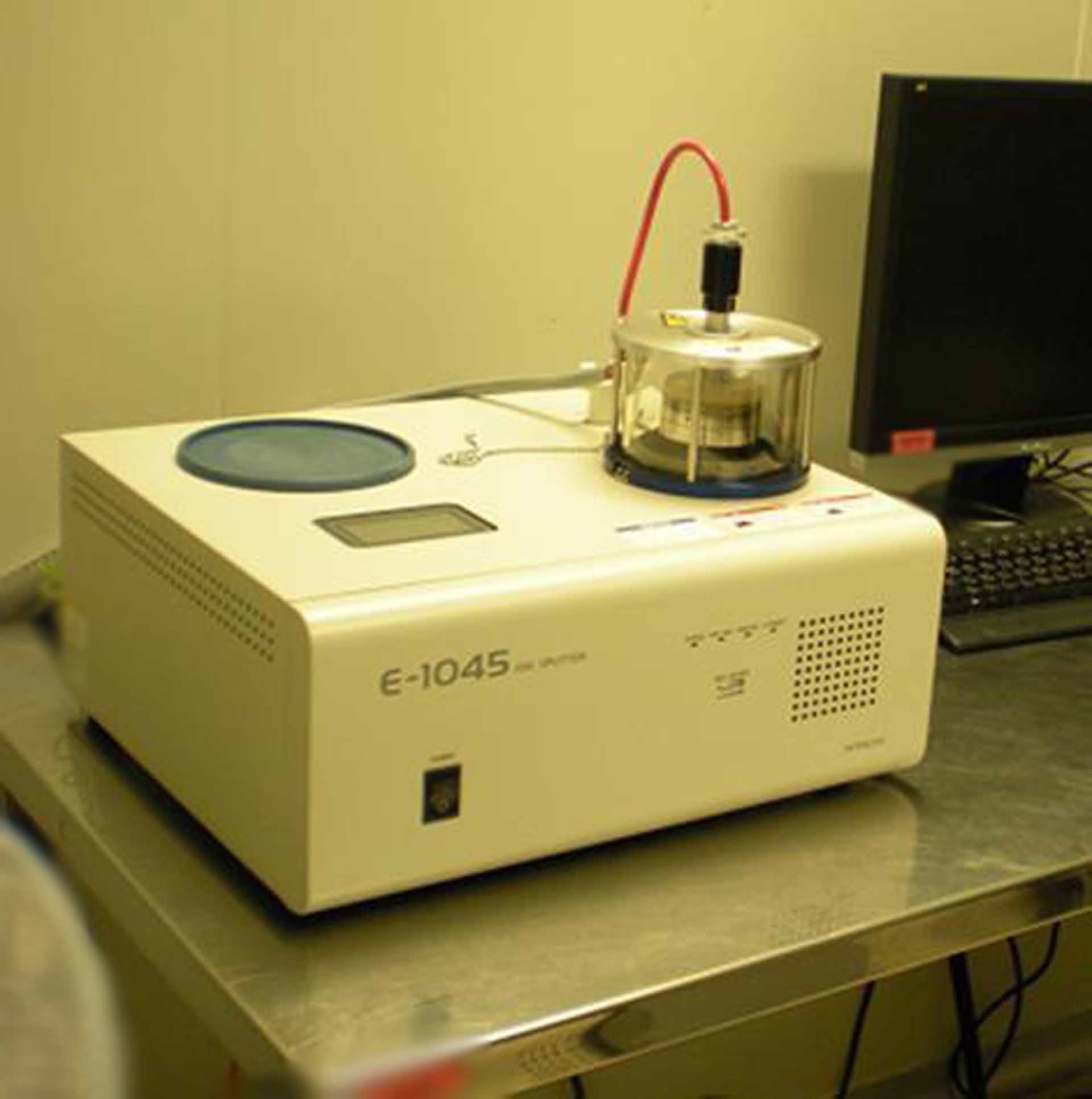
Ion coater
HP Office Jet Pro C8194A Printer
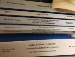
Manuals included
4 kVA For voltage other than 100V AC
Power requirements: 100V AC (±10% ), Single phase, 50/60 Hz
2008 vintage.
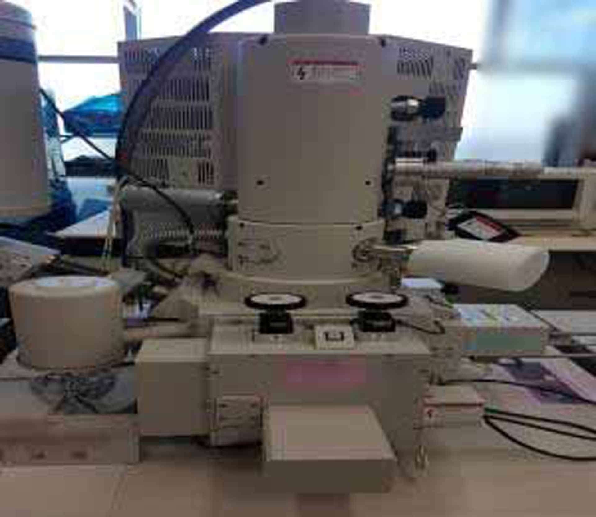
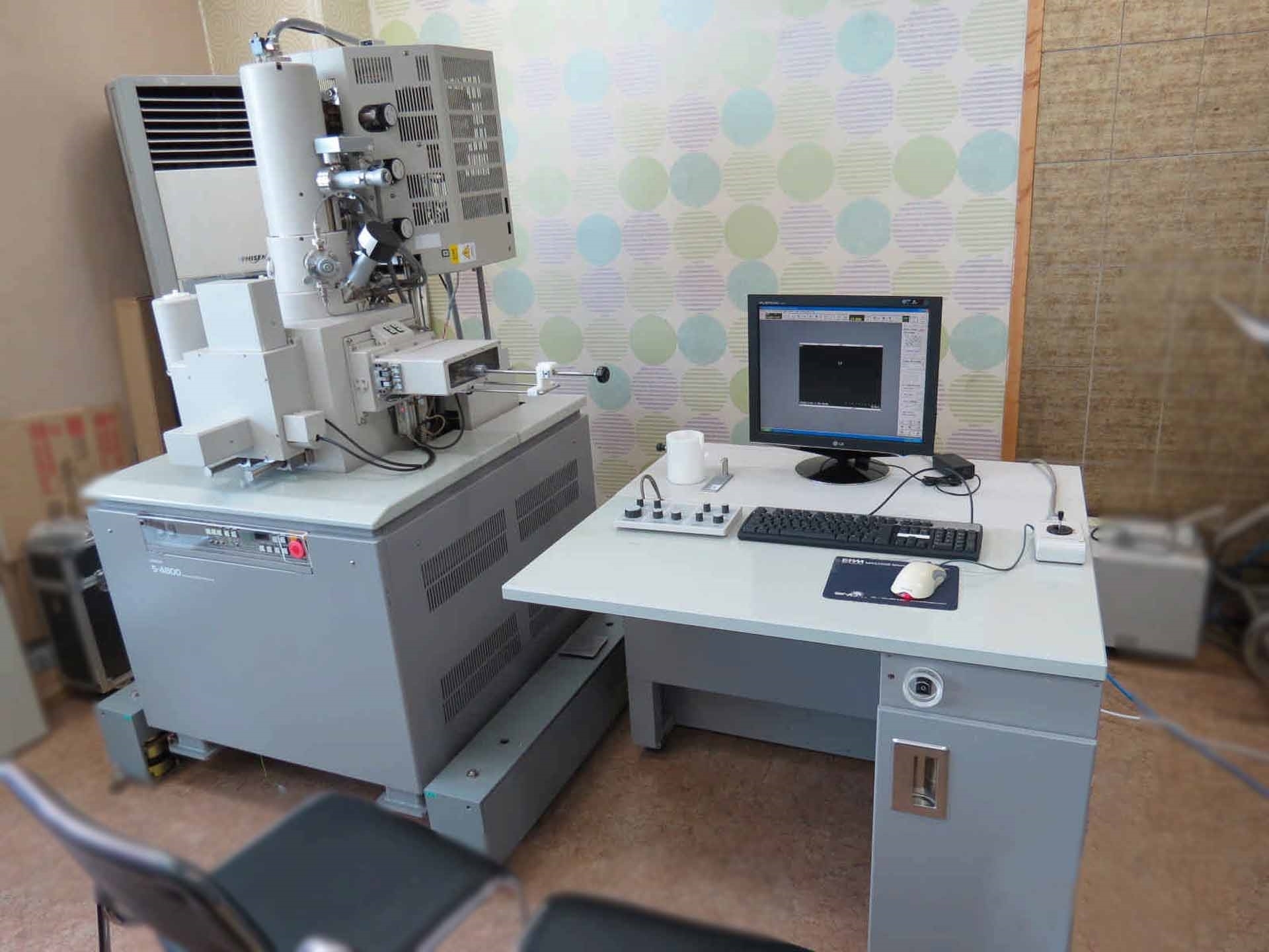
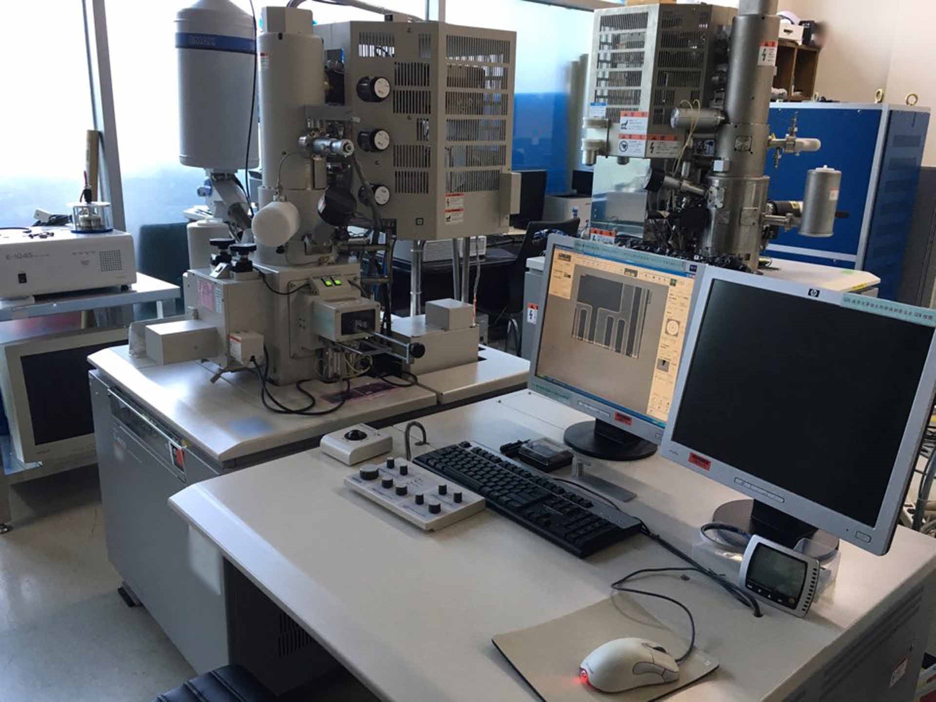
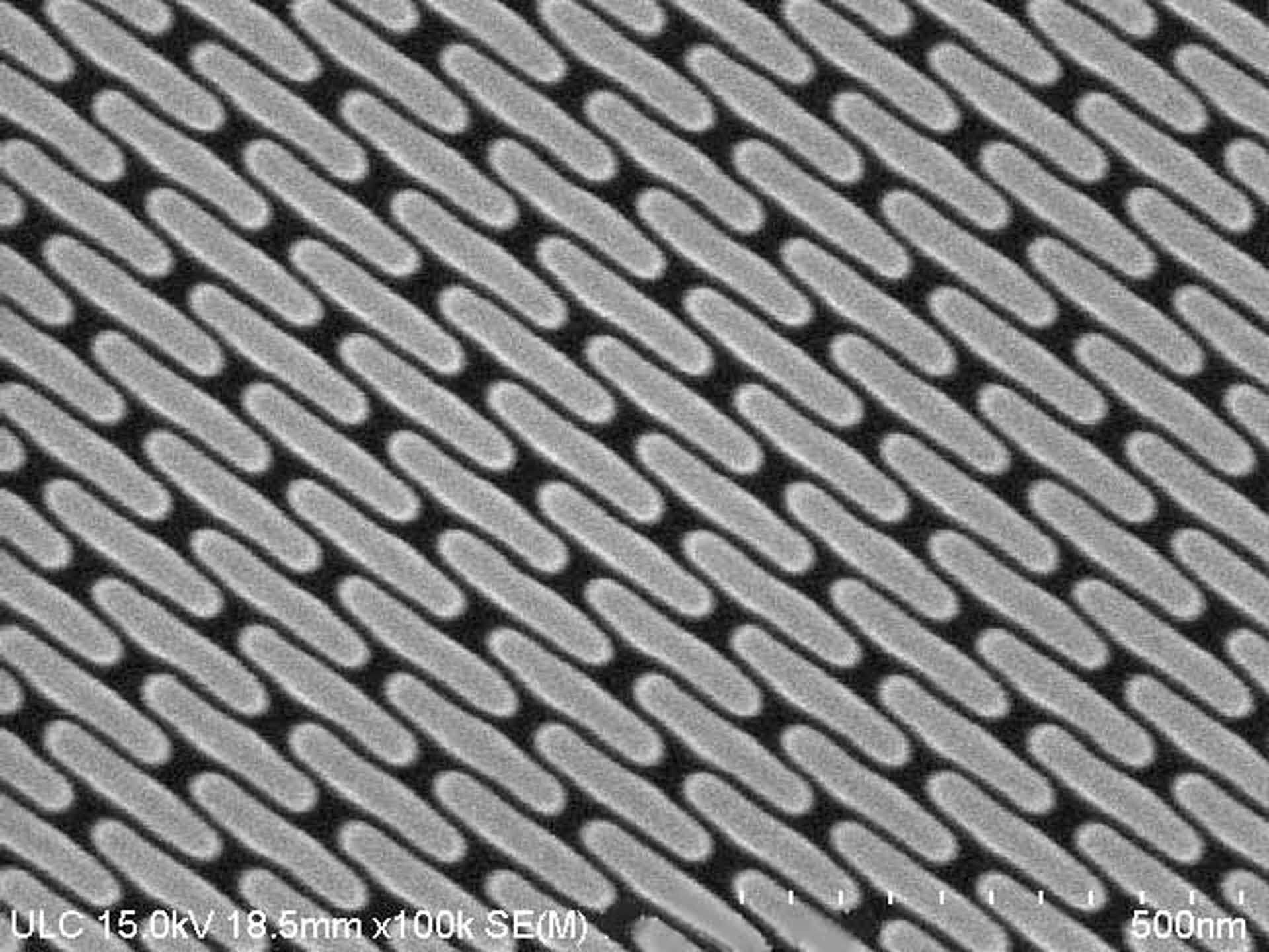
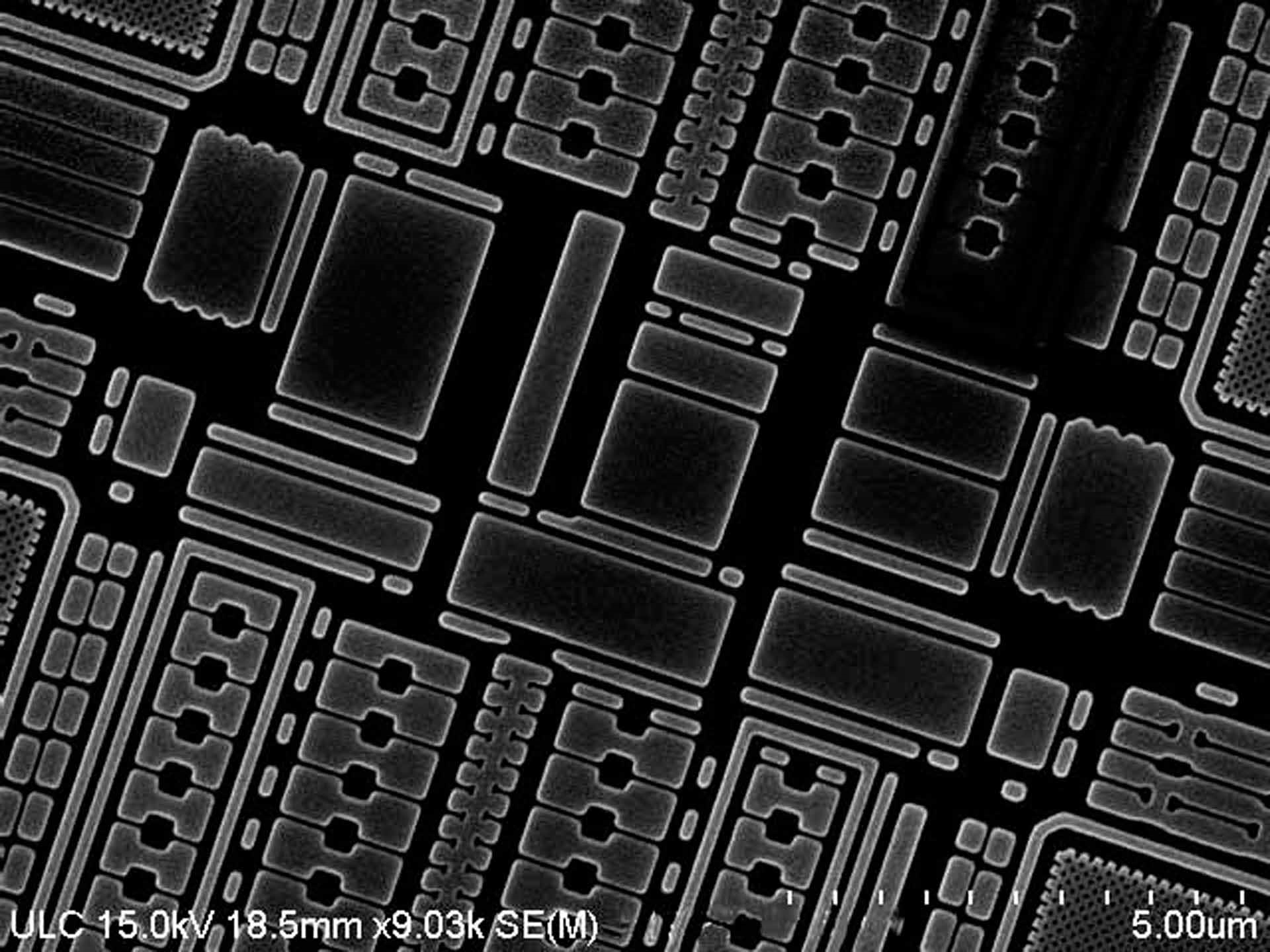
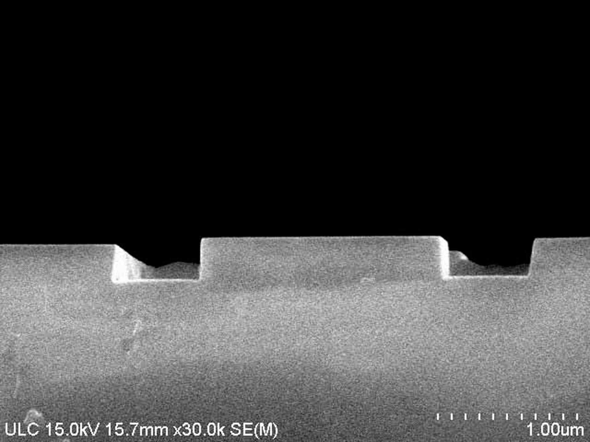
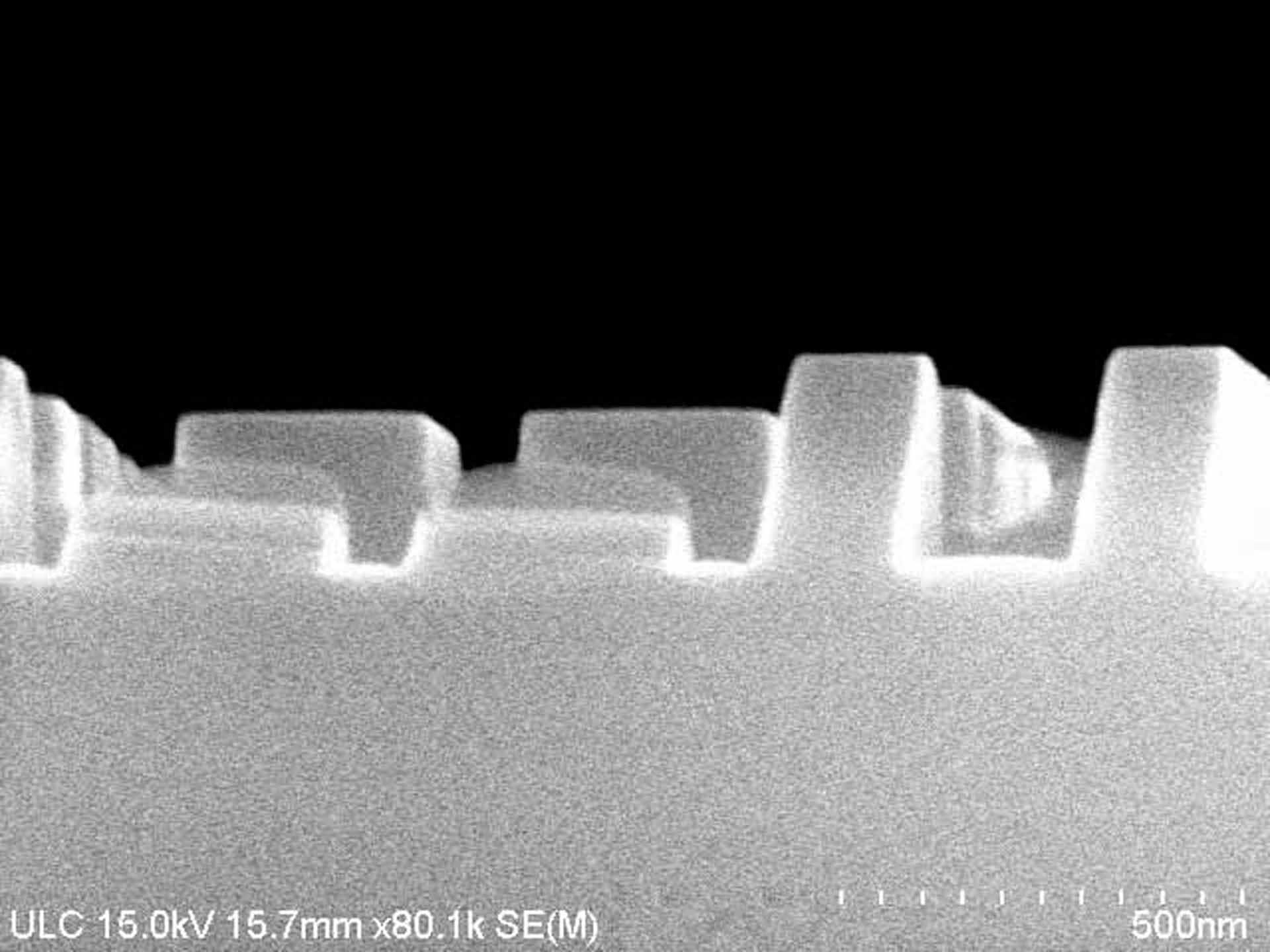
HITACHI S-4800 is a scanning electron microscope (SEM) that provides both excellent image quality and versatile analysis capabilities. This high-end instrument is designed for a range of research and industrial applications, such as materials and life science investigation, specimen analysis, failure analysis, cross-sectioning and surface measurements. HITACHI S 4800 is equipped with a tungsten-filament electron emitter, variable pressure (VP) and variable pressure variable temperature (VPVT) functionalities. The VP and VPVT modes enable specimens to be viewed at different pressure levels and temperatures, allowing for deeper and more accurate characterizations. In addition, the microscope features a large detected area of 140mm in diameter and has high resolution, up to 1nm point resolution in secondary electron image (SEI) mode. Furthermore, the advanced low vacuum mode (ALV) offers the capability of detecting SEM Image on non-conductive materials. The flexible design of S-4800 enables it to be used for a variety of techniques, such as backscattered electron (BSE) imaging, EDX elemental analysis, and WDS for qualitative and quantitative elemental analysis. Additionally, the stereo and 3D imaging capabilities provide a comprehensive characterization of specimens. The advanced imaging includes live and monochrome displays, as well as digital image measurement tools to enable precise measurements and designations of internal structure and surface properties. Additionally, the large sample chamber and high lift stages enable efficient sample handling and manipulation. S 4800 also has a wide range of accessories, such as an array of advanced detectors and electron column upgrades, and an automatic imaging system that enables automated acquisition of images. Moreover, advanced energy filtering aids the study of energy-dispersive X-ray signals. Overall, HITACHI S-4800 is a powerful, high-end SEM designed to meet the demands of various research and industrial applications. It offers excellent image quality and advanced analysis capabilities via its versatile design, enabling users to get precise and accurate view of the specimen being analyzed.
There are no reviews yet
