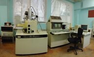
Used HITACHI S-806 #293634215 for sale
URL successfully copied!
Tap to zoom
















ID: 293634215
Field Emission Scanning Electron Microscopes (FE-SEM)
Maximum specimen size, 6"
Secondary electron image
Resolution: 6 nm (60 A) - 25 kV
Magnification: 50x - 100,000x
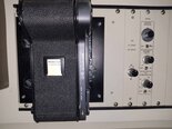
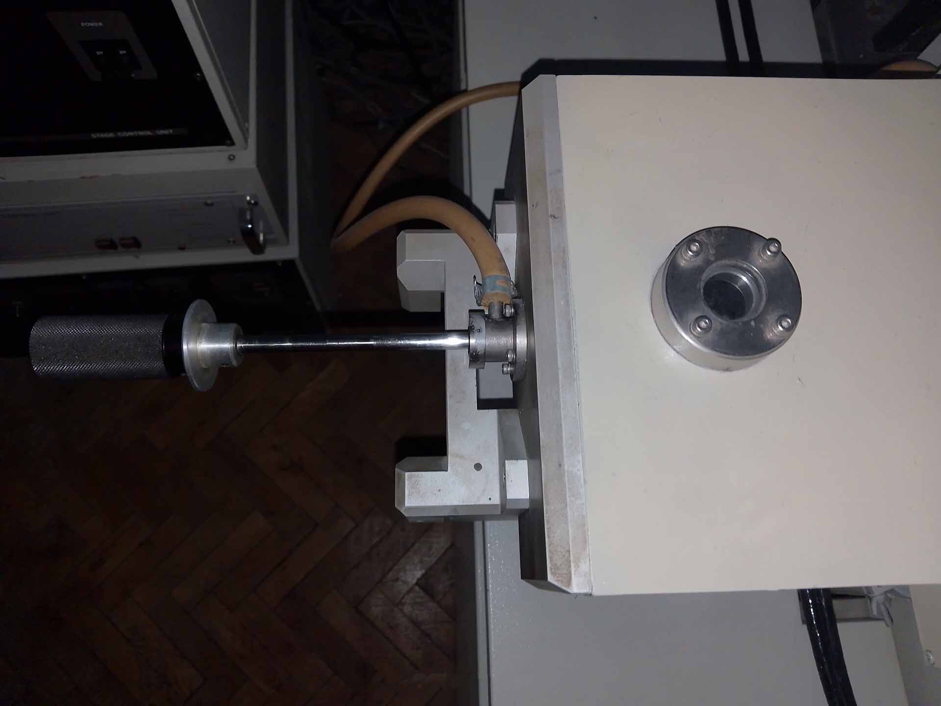
Manual loader, 6"
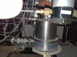
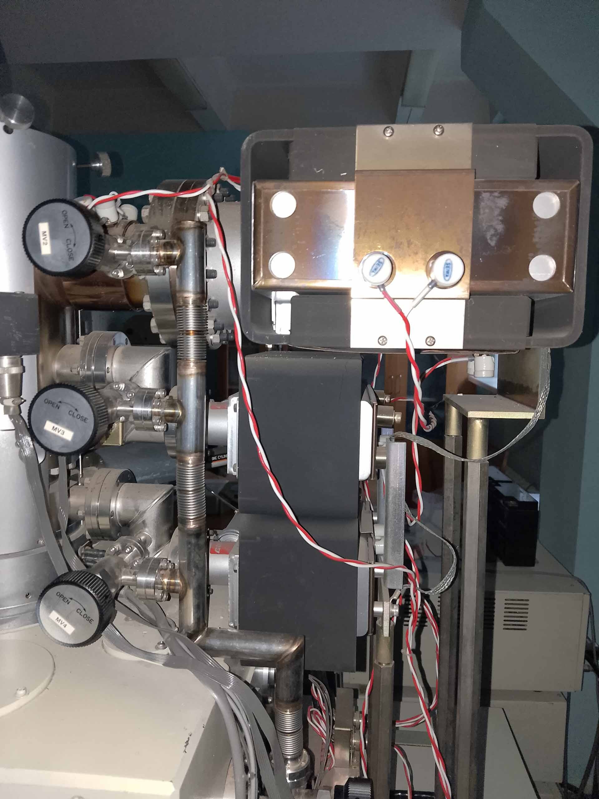
(3) Ion pumps
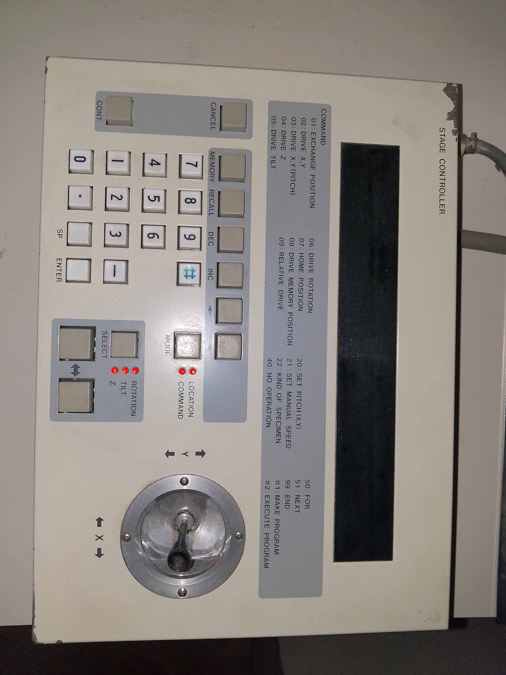
Stage controller
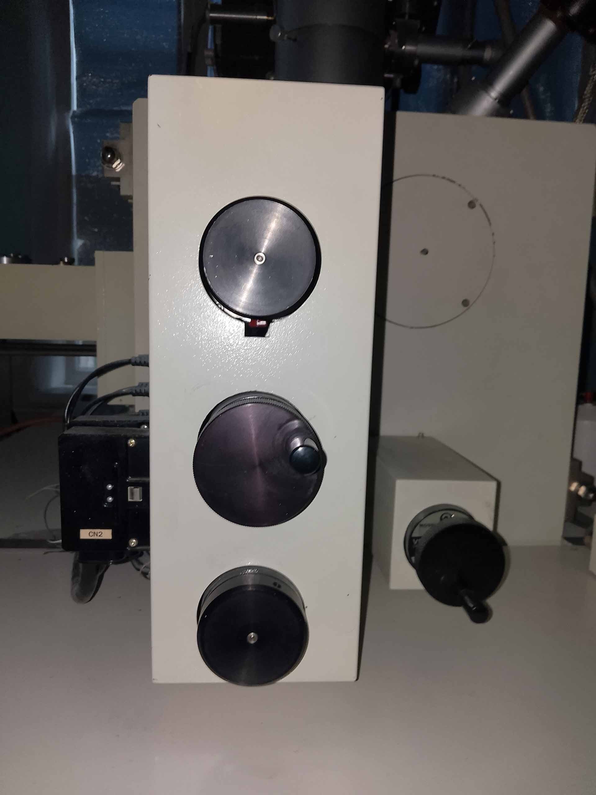
Detecter
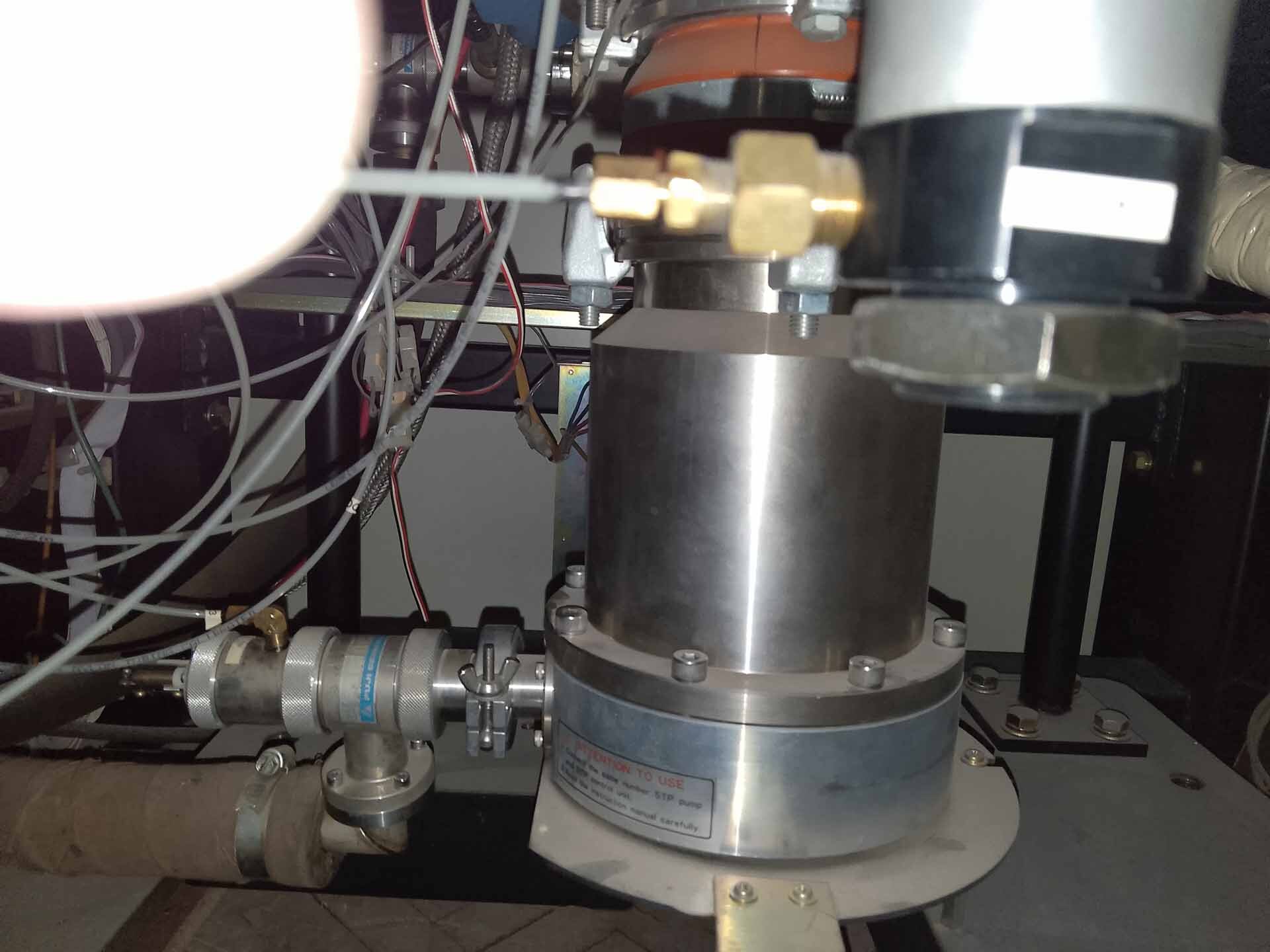
Turbomolecular pump
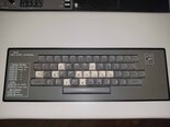
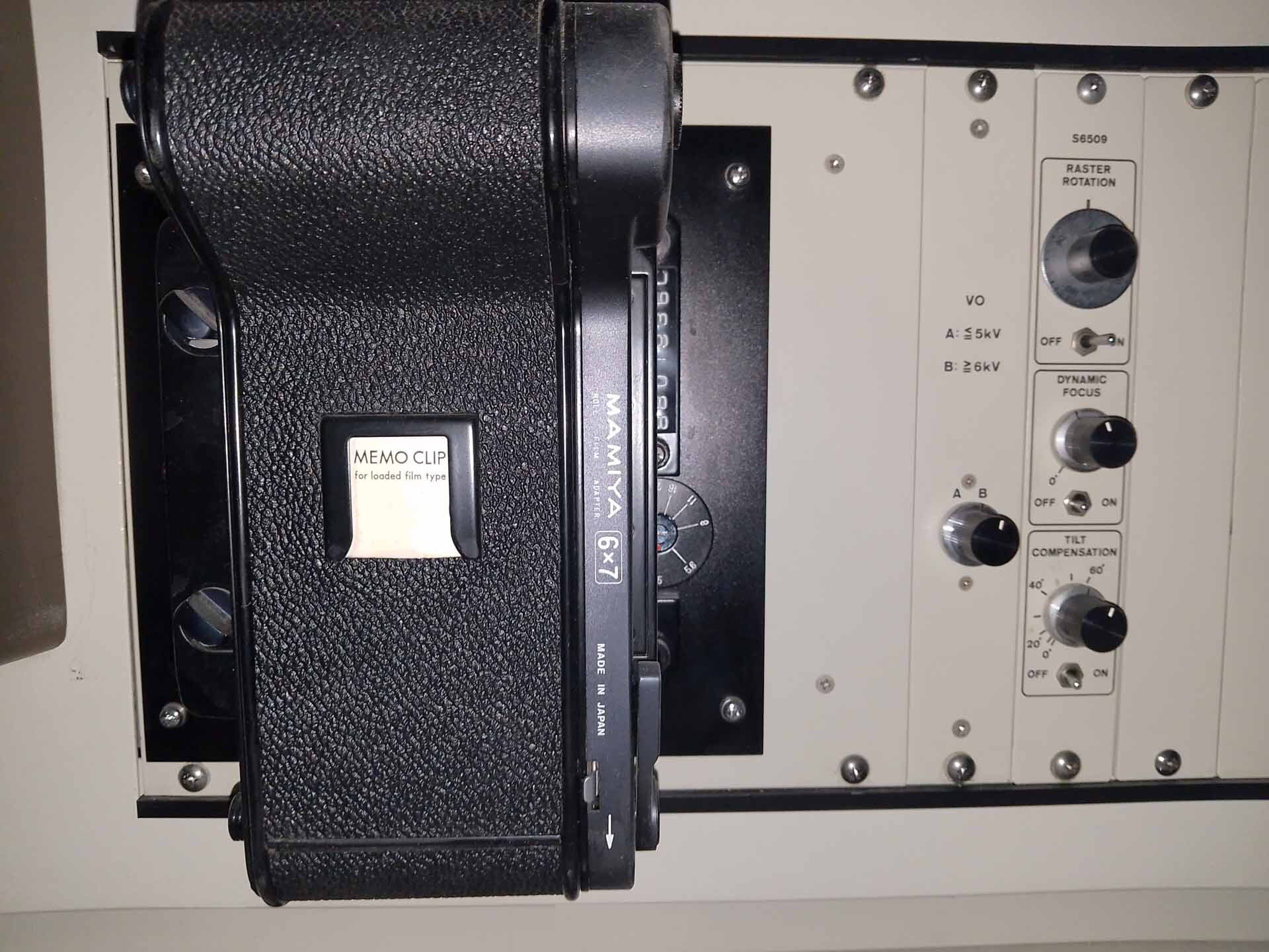
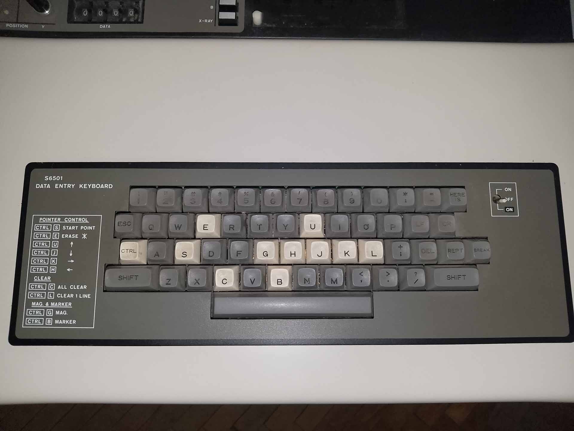
Keyboard
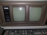
Display Unit
Specimen stage:
Movement range:
X and Y: 0 to 150 mm
Stage drive: Pulse motor control
Joystick control (Auto eucentric)
Z (WD): 5 - 35 mm
Rotation: 360°
Tilt: 0 - 60°
Electron optics:
Electron gun: Cold field emission type
Accelerating voltage: 0.5 - 5 kV (variable in 0.1 kV step) / 5 - 25 kV (variable in 1 kV step)
Emission extracting voltage: 0 - 6.3 kV
Lens system: 2-stage electromagnetic lens
Objective aperture: click-stop type 4 openings, alignable outside column, self-cleaning type thin aperture
Scanning coil: 2-stage electromagnetic type
Stigmator: 8-pole electromagnetic type (X, Y)
Control and display system:
Scanning modes
Viewing CRT
Digital image registration computer system.
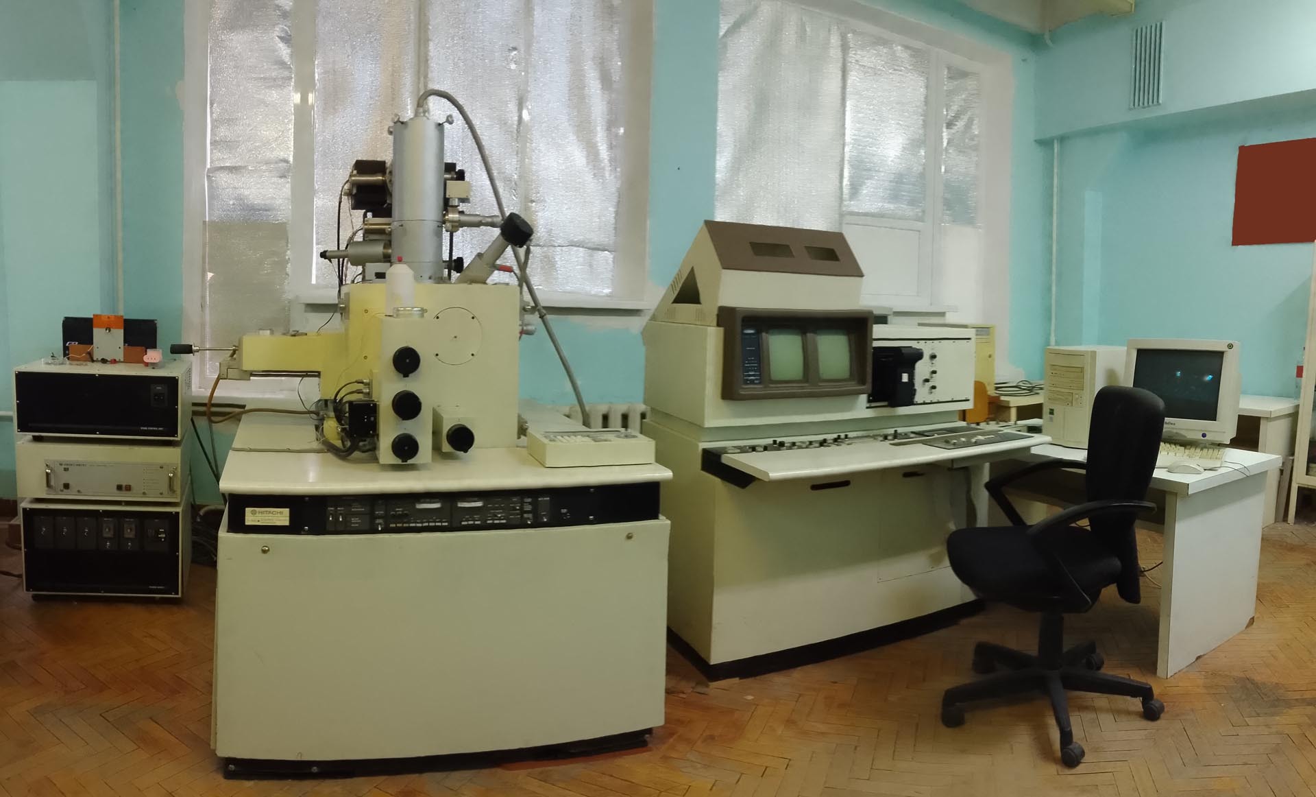
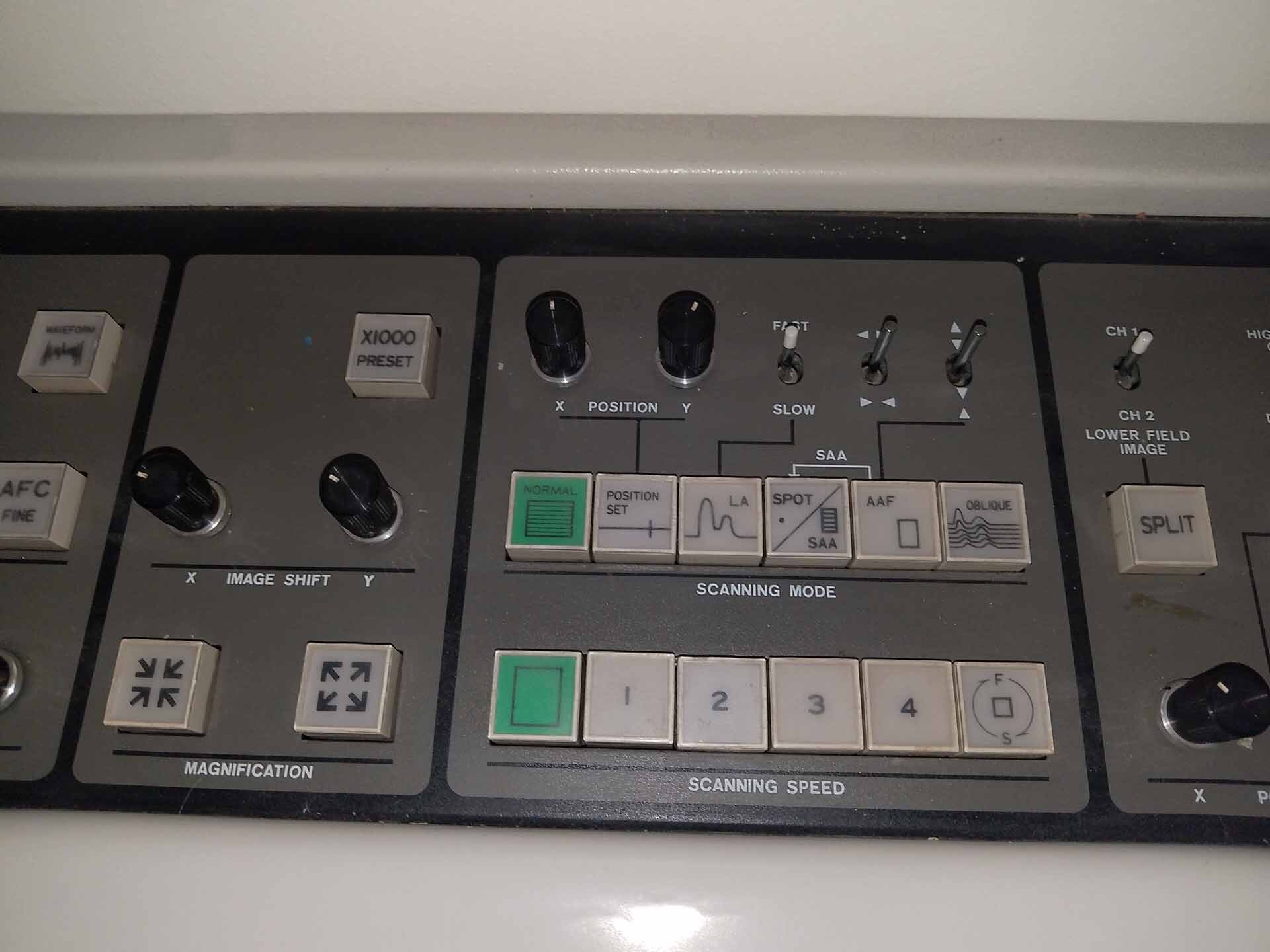
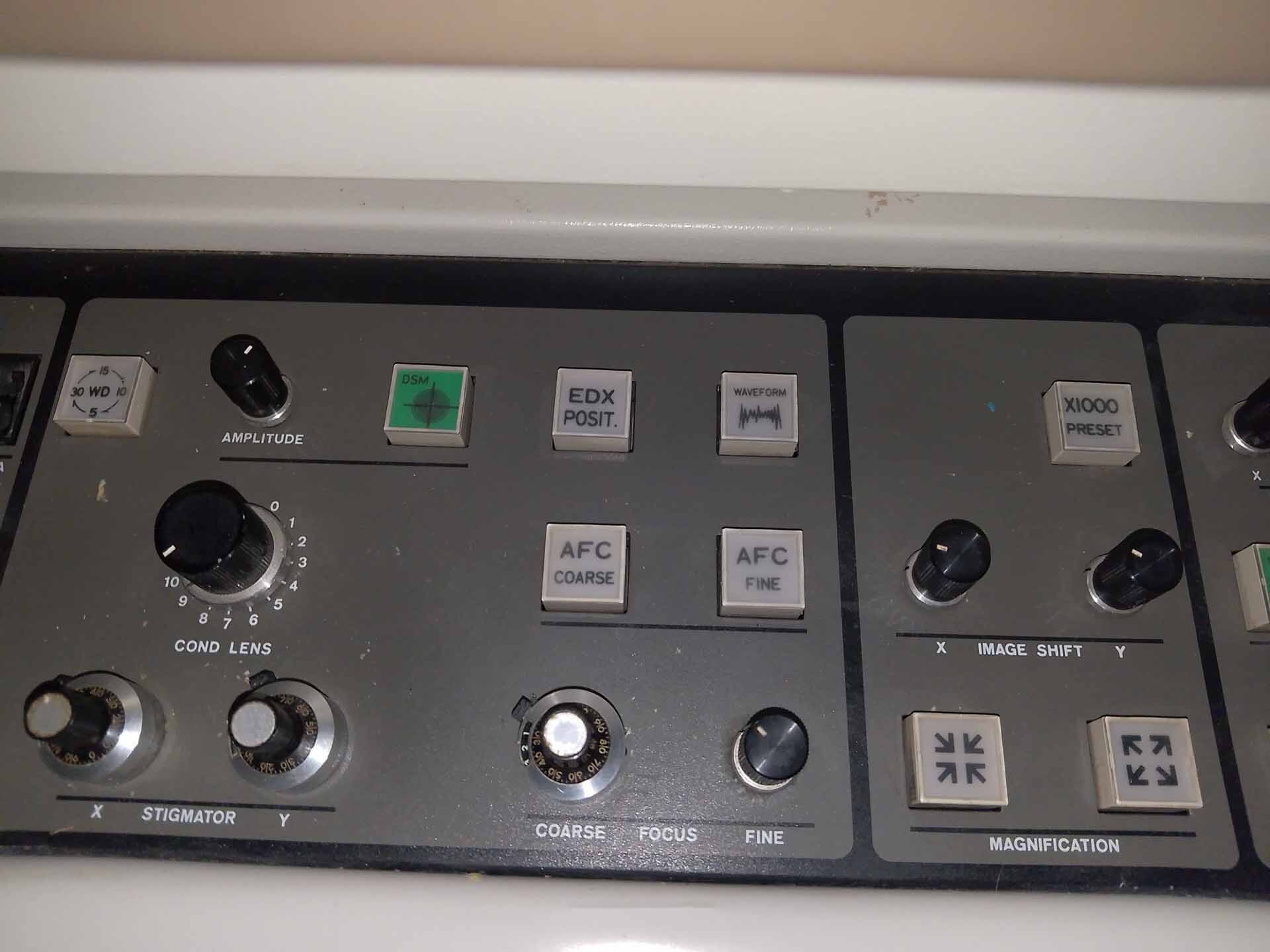
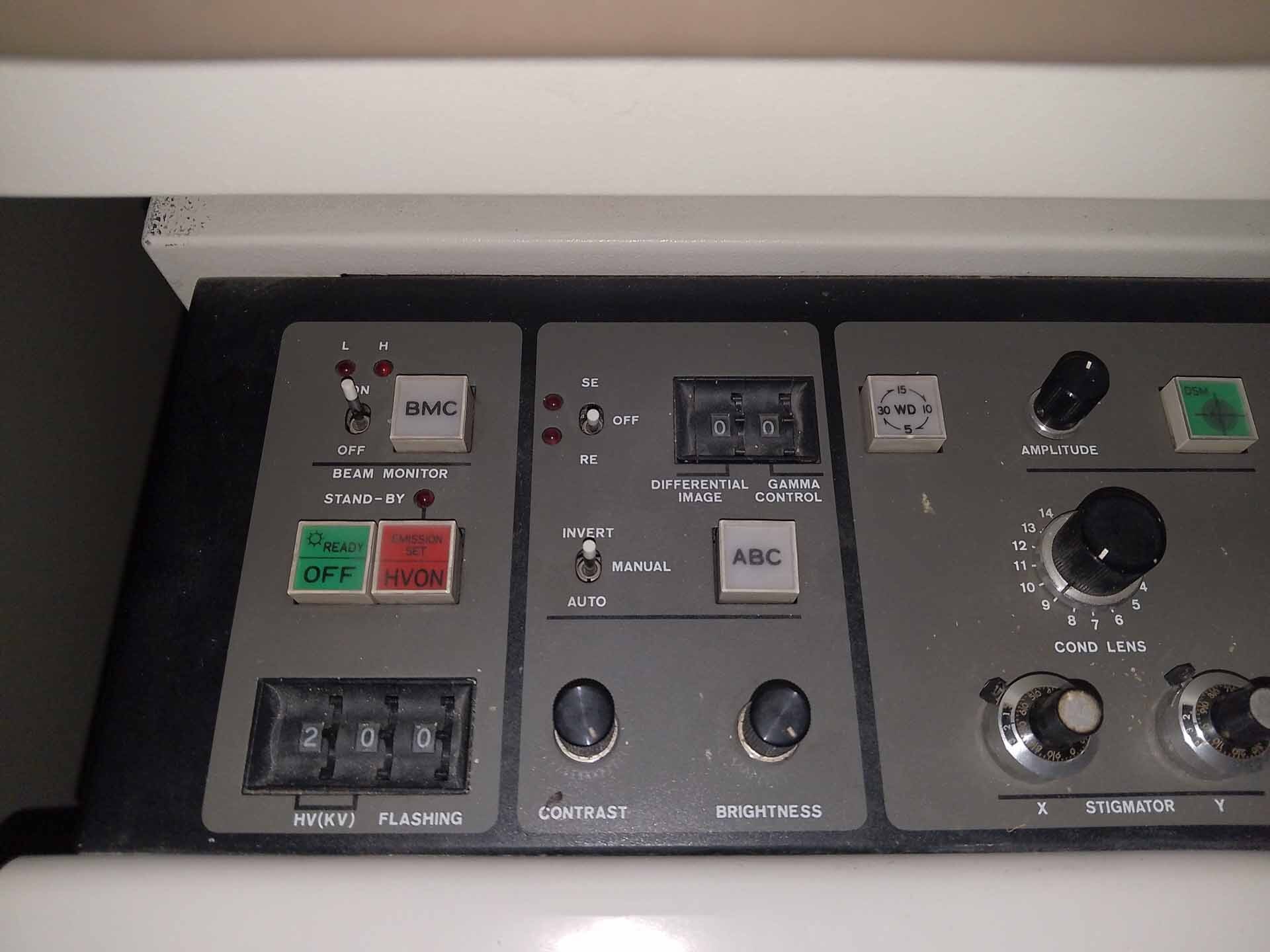
HITACHI S-806 Scanning Electron Microscope (SEM) is a multi-mode, high-performance SEM, capable of viewing specimens at high magnifications with nanometer level resolution. HITACHI S806 is capable of producing secondary electron, backscatter electron, and Auger electron images, as well as X-ray spectra, chemical state maps, and other elemental characterization data. S 806 features a field emission gun (FEG) source, providing excellent beam stability and minimizing changes due to gas conditions, allowing for greater precision and measurement results. The FEG source has a variable accelerating voltage of 0.5 up to 30 kV, with an operating range of 1-25 kV. To maximize viewing and imaging abilities for samples, HITACHI S 806 is equipped with various commands for adjusting scan size, resolution, magnification, and contrast. The SEM sample chamber is large enough for samples up to 100 x 100 mm, and an environmental chamber is available for controlling the atmosphere and pressure. S806 also uses a 12-bit SEM imaging detector, with a pixel size of 5 x 5 microns, to produce images with high levels of detail. To ensure precise results, S-806 is capable of integrating a variety of detectors with the SEM system. These detectors include power spectrometers, detectors for composite analysis of composites and semiquantitative analysis, and optical detectors for determining the height of samples. HITACHI S-806 can be used in conjunction with a conventional stereo microscope to capture 3D images. It also supports a variety of automated stage options to simplify specimen loading, positioning, and measurement. Overall, the advanced features of HITACHI S806 Scanning Electron Microscope make it an ideal choice for nanometer scale imaging and characterization. Its versatile and precise capabilities, FEG source, automated stage support, and various detection systems allow for accurate results in a wide range of scientific applications.
There are no reviews yet
