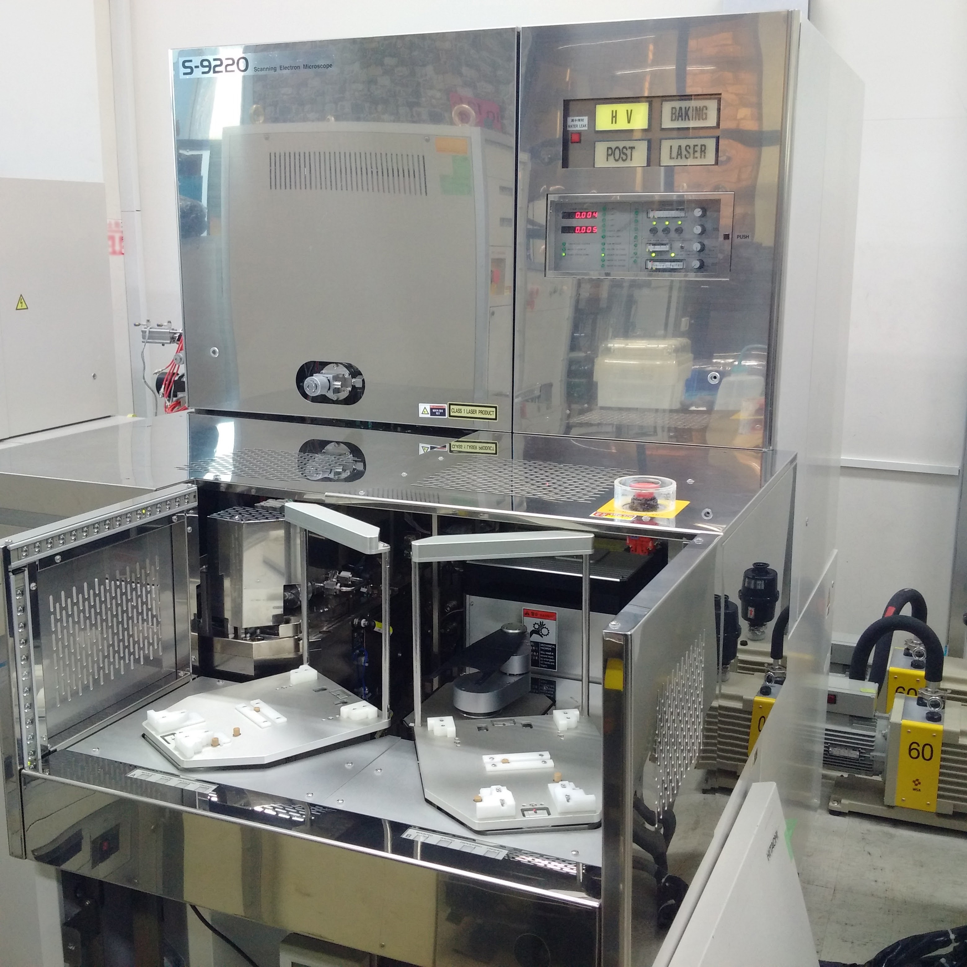
Used HITACHI S-9220 #9200635 for sale
It looks like this item has already been sold. Check similar products below or contact us and our experienced team will find it for you.
Tap to zoom


Sold
ID: 9200635
CD Scanning electron microscope, 8"
System information:
Work station
Model: B180L
O/S: HP Unix
General specifications:
SEMI/JEIDA standard orientation
Flat or V notch wafers
Measurement method: Cursor / Line profile
Measurement range: 0.1µm ~ 2.0 µm
Measurement repeatability: ±1% / 3 nm (3 sigma) whichever larger
Throughput: 56 Wafers/hr
Measured points: 1 Point/chip, 5 Chips/wafer
Secondary electron image resolution: 3nm
Image magnification: 500 ~ 300kx
Electron optics system:
Electron gun: Schottky emission
Accelerating voltage: 500 ~ 1,600 V
Lens system:
Scintillator/photomultiplier detection system
ExB Filter
SE / BSE Electrons
Objective lens aperture: Heating type movable aperture
Fine adjustment possible
Scanning coil: 2 stage electromagnetic deflection
Stigmator coil: 8-pole electromagnetic type (X, Y axes)
Probe current monitoring: Faraday cup incorporated, with automatic measurement
Optical microscope: 1.2-mm-square visual field, monochrome image
Stage:
Movement range: X, Y: 0 ~ 200mm
Driving method: Pulse motor
Control/speed:
Positioning contro
Linear encoder
Maximum speed: 100mm/s
Loader:
Wafer transfer:
Cassette to loader chamber: Auto transfer via wafer transfer robot
Loader chamber to stage: Auto evacuation and auto loading
Wafer transfer robot system:
Random access using two cassettes
Wafer detection in cassette: Automatic detection via wafer searcher
Chucking method: Vacuum chucking on back of wafer
Orientation flat / V notch detection: Non-contact auto detection via optical sensor
Control and display system:
CRT:
EWS 21 Type monitor display of SEM and OM images
GUI Operation screen
Wafer map
Measured values
Stage coordinates
Scanning modes:
TV Scan
HR Scan
Slow scan
With auto brightness/contrast function
Image Processing:
Hardware processing using DSP (Option)
Recording:
Video printer output function (Option)
Image filing function (Option)
Safety device: Equipped with emergency off swith
CD Measurement data processing system:
File storage: Storage function for various setting parameters, measurement results
Storage media:
Hard disk (9.1GB) incorporated in EWS
3.5 Type magneto-optic disk
3.5" Floppy disk
Data process function:
Statistics output
Worksheet system
Output of measured values in real time graph
Printout: 80 Character thermal printer
Evacuation system:
Full automatic
Dry / Clean evacuation
Vacuum pumps :
(3) Ion pumps
(2) Turbo molecular pumps
(2) Oil rotary pumps
Safety devices:
Protective device against:
Power failure
Column vacuum level drop
Dry air pressure drop
Cooling water flow rate drop
Grounding: 1000 or less (Single)
Nitrogen:
200 ~ 680kPa
Pipe outer diameter: 6mm
Vacuum:
P: 1.3 ~ 21.3 kPa or less
Pipe outer diameter: 6mm
Circulation cooling water:
98.1 ~ 196kPa
Pipe outer diameter: 15mm
Environmental conditions:
Magnetic field:
AC magnetic field: 0.3µT or less
DC magnetic field: 0.1µT or less
Vibration:
Horizontal direction:
1Hz: 3µm (P-P) Max
2Hz: 0.7µm (P-P) Max
3Hz: 1.2µm (P-P) Max
4Hz: 2µm (P-P) Max
5Hz: 3µm (P-P) Max
10Hz: 3.5µm (P-P) Max
Noise: Less than 75 dB
Room temperature: 20 ~ 25°C (Δt = ±2°C)
Humidity: 60% or less
Utility:
Power:
100 V, 200 V, 208 V, 230 VAC
6 kVA
50/60 Hz
1 Phase
2001 vintage.
HITACHI S-9220 is a state-of-the-art scanning electron microscope (SEM). This electron probe microscope is a versatile platform for analyzing structures down to sub-nanometer resolutions. It offers excellent pixel resolution in the 1-2 nanometer range and optimizes image quality and resolution across the length and breadth of the sample being examined. The high magnification of HITACHI S9220 up to 1,000,000x magnitude enables researchers to carefully inspect the tiniest details of the nanoscale objects. The conventional SEM requires a vacuum chamber for operation and S 9220 is no exception; however, it uses a large commercial dry vacuum equipment which ensures a high degree of reliability while offering superior performance. This vacuum system limits the effects of ions, air molecules, and other particles that may cause artifacts to be present in the image. The use of an Ultra Low Vacuum (ULV) unit keeps the pressure in the chamber constant for a longer period of time, allowing for the collection of more information from the sample and producing better image quality. HITACHI S 9220 is equipped with an imaging machine that includes a field emission gun to emit electrons at high-energy level, a condenser lens to focus those electrons onto the sample, a detector to collect the signals and an image detector to process the image. This imaging tool provides superior performance due to its precision lenses and high-energy electrons that can easily penetrate the sample and map its atomic structure. The sample-stage of S-9220 is able to move the specimen around 4-axes (x, y, z and rotation) to precisely manipulate the required area that needs to be imaged. The specimen holder allows for specimen's that range from large and heavy samples to delicate and small samples. In addition, the sample stage also features a temperature-controlled unit to contain the sample during cooling and analyze its chemical composition. S9220 also incorporates two measurement tools to analyze the sample. The first tool is an energy dispersive x-ray spectray (EDS) detector. This unit enables the chemical composition of the specimen to be determined with very high accuracy. The other tool is an x-ray diffraction tool (XRD). This gauge allows researchers to determine the crystalline structure of materials from various angles and orientations. Overall, HITACHI S-9220 is the ideal scanning electron microscope for conducting detailed analysis of nanoscale objects. Its large commercial dry vacuum asset, high-energy electron imaging model, and multiple stages and tools make this SEM reliable and precise, allowing researchers to obtain accurate and high-resolution images with ease.
There are no reviews yet