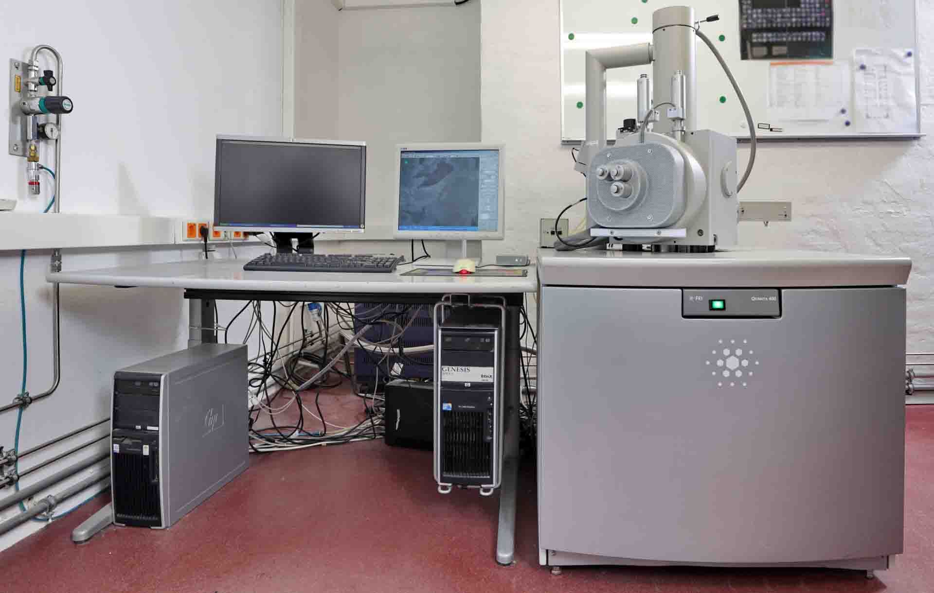
Used PHILIPS / FEI Quanta 400 #293585905 for sale
It looks like this item has already been sold. Check similar products below or contact us and our experienced team will find it for you.
Tap to zoom


Sold
ID: 293585905
Scanning Electron Microscope (SEM)
Dispersive X-Ray detector
Ultra-thin detector window
Backscatter electron detector
USB controller:
DC-Isolated USB 2.0 Interface
HV-VAC Controller:
DAC Uni / Bipolar: ± 10 V (8 x 16 bit)
ADC for emission (5 x 10 bit)
VAC
Operating voltages
(16) Digital inputs and outputs
Security link to vacuum system
Control power supply
Turn-on of power supply
Emergency turn-off of electronics at malfunction
Module for deflection coils:
(2) Single deflection systems
(4) Double deflection systems
Magnification: 4 step and 16 bit
Scan rotation
Scan shift fine
Correction of tilt
Orthogonality
Rotation offset
Module with bipolar power supplies:
DAC for current/voltage supplies (8 x 16 bit)
Beam shift
Tilt coils
Image shift coils
Stigmator coils (8 pole)
Filament imaging
Stigmator image
Power supply: 4 x ±500 mA, 4 x ±10 V
Module for objective lens and condenser lenses:
Unipolar power supply
Objective lens: 2 x 16 bit DAC (coarse, fine), maximum 6.5 A
(2) Condenser lenses: 16 bit DAC, maximum 6.5 A
Image system:
Maximum pixels: 16384 x 16384
Pixel clock: 200 ns
D/A Converter for analog input signals (4 x 12 bit)
Counter for mapping (12 x 16 bit)
Simultaneous acquisition
(4) Analogs
(12) Digital input signals
Image acquisition
Windows 2000: Up to 10 (x86, x64)
Full screen mode
Slow scan
Mapping
Oversampling for noiseless images: Up to 32000
Line averaging
Frame averaging
Reduced area scan
ROI Scan
Qualitative and quantitative with EDS/WDS
AVI Function
Signal monitor to control image signals
Trigger inputs and clock outputs for point
Mains synchronization for slow scan
Thumbnail bar for acquired images
Image processing:
Image browser
Loading and saving of images file types: TIFF, BMP, JPEG, PNG, GIF
Auto save function
Creation of image sections
Image rotation
Image labeling functions
SEM Parameters
Power supply:
Analog power supplies: ±15 V, +5 V, +12 V
Switching power supply for high power objective and condenser lenses
Module for PMTs: 2 x 0 to 1.5 kV
Module for scintillator: 12 kV
Grid voltage: 0 V to 400 V.
PHILIPS / FEI Quanta 400, or Q400 for short, is a scanning electron microscope (SEM) designed for use in materials characterization and forensic analysis. It features a field emission source - a small, localized electron source at the heart of the SEM. This enables the instrument to collect high-resolution electron images, maps, and spectra on a wide range of sample types. The Q400 is a solid-state system and therefore does not require liquid nitrogen for cooling. The system features a large digital resolution controller to deliver accurate and precise beam current at the sample. For imaging, the Q400 can deliver high-contrast images with a resolution of up to 0.5nm, enabling accurate determination of surface topography. Additionally, its SEM capabilities can be used to obtain chemical mapping information with EDS (energy dispersive spectroscopy), enabling rapid elemental mapping across the sample surface. To ensure optimal SEM imaging and analysis, the sample must be pre-prepared with the Q400 supplied 'standard dummy' sample holder. This works to reduce or eliminate charging effects on the sample. Investigating the interaction between surfaces can be explored using the latex sphere attachment and crystal orientation can be collected using the goniometer. Below the sample surface, the Q400 can be used to explore local surface structure through the retrieval of a larger area by collecting a series of images. These can then be merged and stitched together. This stitch ability allows researchers to explore wider areas in detail for structural and compositional information. The capture of SEM images coupled with EDS mapping can often spark a number of questions. For example, the average grain size of a specimen, the size and distribution of microstructural constituents or any other morphological or compositional analysis. The Q400 supports many software programs that can answer these questions. FEI Quanta 400 is a versatile and reliable scanning electron microscope with numerous features allowing it to be used for a wide range of applications, from materials analysis to forensic investigation. Its high-resolution imaging, EDS mapping, goniometer attachment and surface imaging capabilities put it ahead of its competitors.
There are no reviews yet