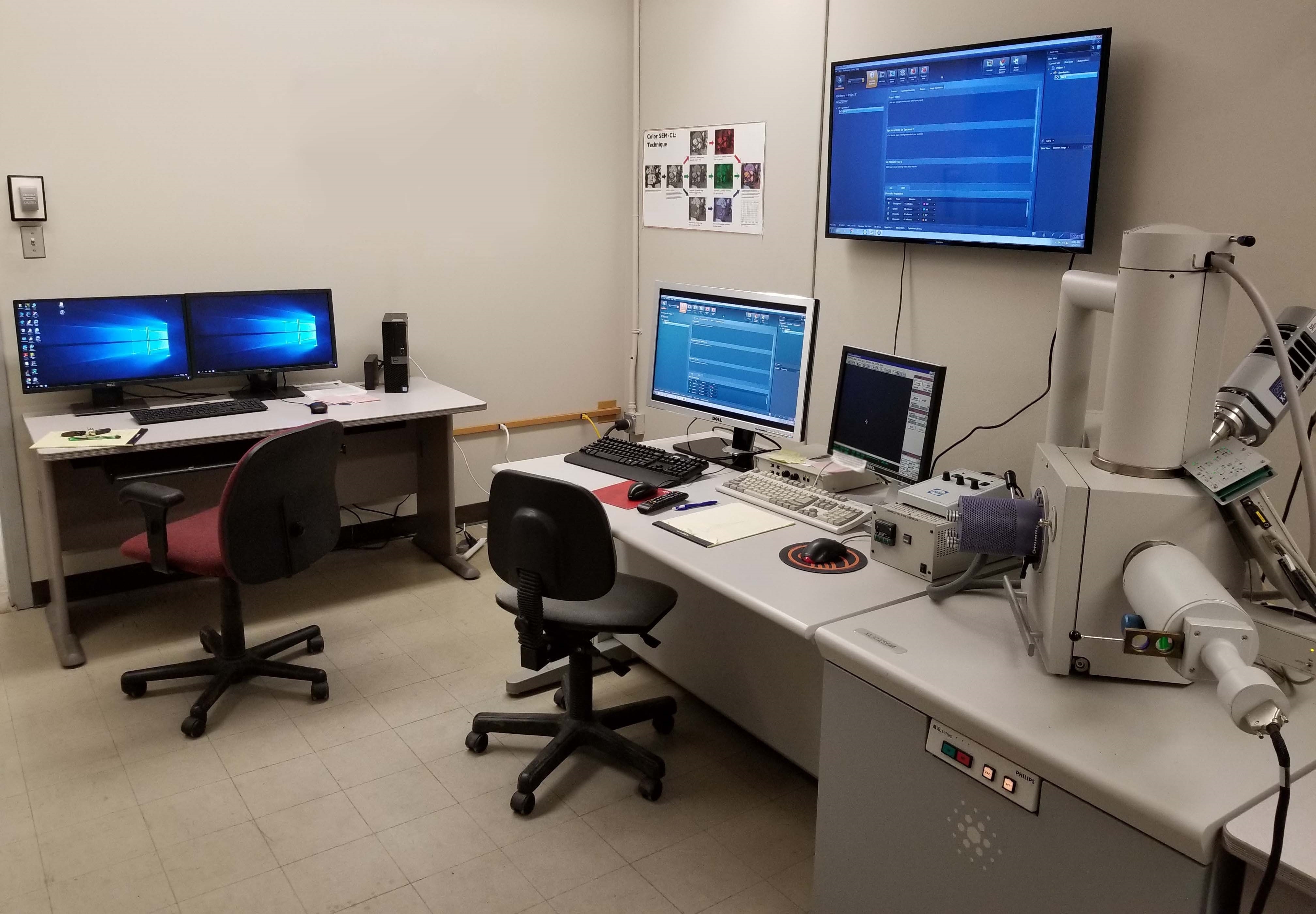
Used PHILIPS / FEI XL 30 #9260099 for sale
It looks like this item has already been sold. Check similar products below or contact us and our experienced team will find it for you.
Tap to zoom


Sold
ID: 9260099
Vintage: 2001
Environmental Scanning Electron Microscope (ESEM)
Magnification: 100x to 100,000x
PELTIER Cooled specimen stage between -5°C and +60°C
Equipped with:
SE and BSE detectors
High and low (ESEM) vacuum modes
Imaging capabilities:
Secondary Electron (SE) detector for imaging under high-vacuum
Backscattered Electron (BSE) detector for imaging under high-vacuum
Large Field Detector (LFD) used between 0.1 and 1.0 torr to detect BSE and SE signals at low accelerating voltage
Gaseous Secondary Electron Detector (GSED) allows SE detection at up to 20 torr
Wide angle GSED allows SE imaging at up to 10 torr
Standard Secondary Electron (ESD) detector contains a 500 µm aperture for imaging up to 20 torr
Standard / Wide angle X-ray ESD has a working distance of 10 mm
Gaseous BackScattered Electron (GBSD) Detector allows BSE, SE, or BSE+SE imaging up to 10 torr using 500 µm aperture
Stage can accommodate
(7) 0.25" SEM stubs
(3) 1" SEM stubs
(2) Standard-sized petrographic thin sections
2001 vintage.
PHILIPS / FEI XL 30 is a type of scanning electron microscope (SEM) that is used extensively in industry, manufacturing and scientific laboratories. It is a powerful instrument that can magnify objects up to 300,000 times or more, allowing users to see intricate details of the sample being examined. It is a dual-beam system meaning that there are two electron beams, one for obtaining high resolution images and the other for beam nanoprobing of the sample. The imaging system is based around a tungsten field emission source that is used to produce the high-energy beam of electrons. These electrons are then focused using a combination of magnets and electrostatic lenses before reaching the sample. The specimen is placed in a vacuum chamber for analysis, where it is viewed at various magnifications on the viewing screen. The SEM uses the secondary electrons from the sample to form an image that can then be analyzed in great detail. The imaging capabilities of FEI XL 30 microscope also include features such as backscatter electron imaging, scanning transmission electron microscopy (STEM), cathodoluminescence and electron beam induced current imaging. This provides researchers with the ability to image and analyze a range of sample characteristics. The electron beamis highly controllable, with a spot size as small as 0.2nm, adjustable beam current and accelerating voltage, and automated sample mapping. The specimen stage has three axes of motion, allowing for accurate positioning, and the high magnification capabilities are enhanced by the microscope's digital readout. Additionally, a variety of detectors are available for electron analysis, including energy dispersive X-rays (EDX) and wavelength dispersive X-rays (WDS). PHILIPS XL30 is a reliable and sophisticated instrument that is capable of producing detailed images and analysis of a wide range of materials and components. Its robustness and range of features make PHILIPS XL 30 an excellent choice for electron microscopy applications.
There are no reviews yet