Used PHILIPS / FEI XL 30 #9300327 for sale
URL successfully copied!
Tap to zoom





ID: 9300327
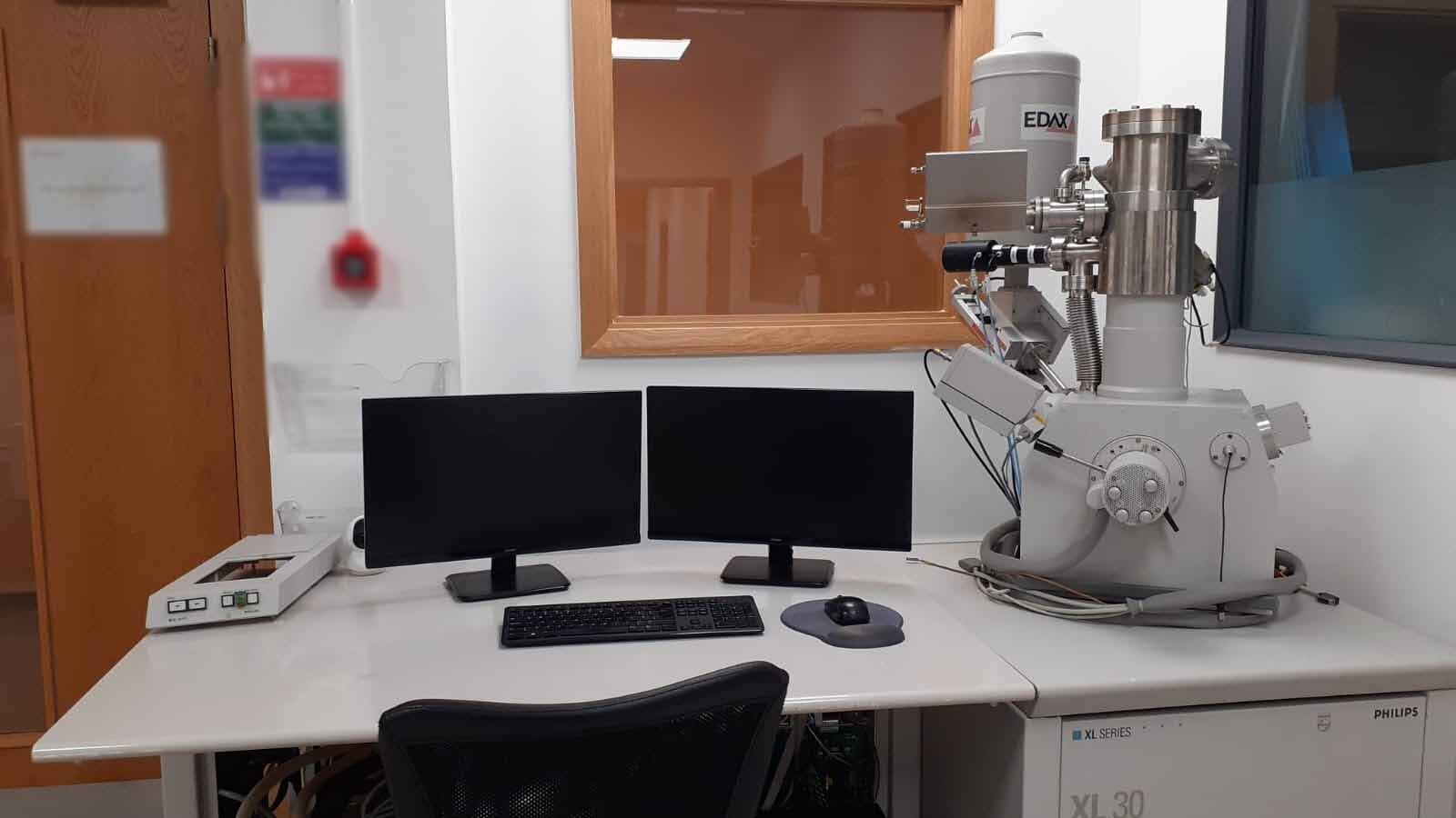
Thermal Field Emission Microscope (FEM)
EDX
BSD
Larger chamber
Chamber camera system
Resolution: 1 nm.
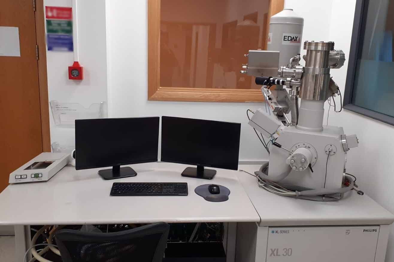
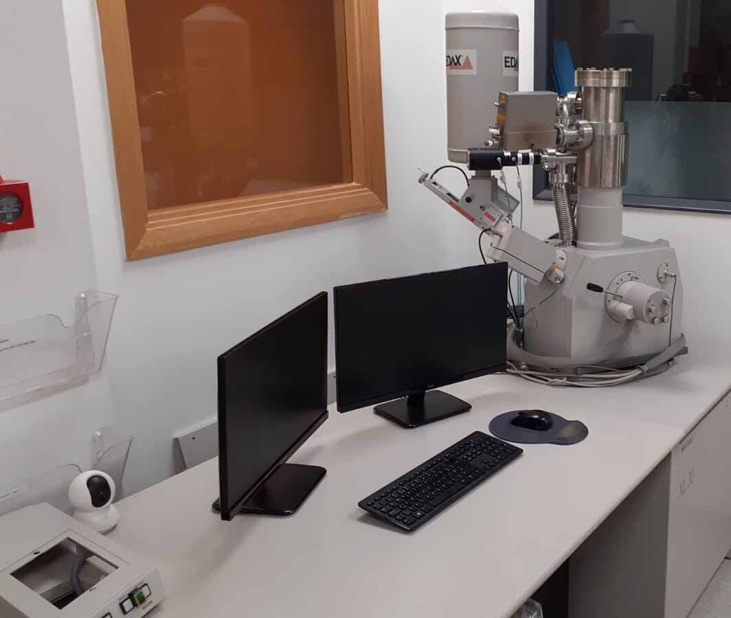
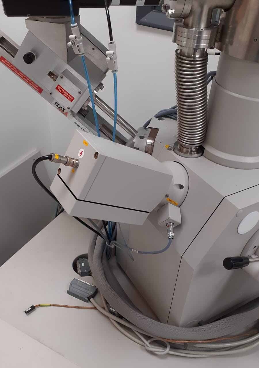
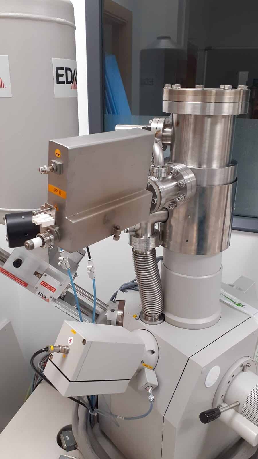
PHILIPS / FEI XL 30 Scanning Electron Microscope (SEM) is a powerful research tool designed for analysis of the topographical, electrical, and chemical properties of a wide variety of samples. FEI XL 30 is designed for use in analytical research, industry and educational settings. The central component of the microscope is the electron column, which contains the lenses, deflectors and magnetic fields that control the beam and produce the images. The column of PHILIPS XL30 is capable of producing a focused beam with a spot size of less than one nanometers in diameter. The electron beam is generated by an electron source, and directed by a series of apertures, lenses and deflectors that exist within the column. This allows the microscope to focus and image at a wide range of magnifications, from 0.3 nm to 500 nm. The samples are viewed inside the vacuum chamber. In order to protect the samples and the column components, a pressure of 5 x 10⁻⁷ mbar must be maintained in the chamber. The specimens are mounted on an electrically-conductive sample holder, which is placed into the chamber. The SEM imaging system has the capability to detect secondary and backscattered electrons, as well as high resolution diffraction. The main imaging modes offered by XL30 are high resolution secondary electron mode, combined secondary and backscattered electron mode, low vacuum mode and auger electron spectroscopy. The microscope also has a full array of spectroscopic capabilities for analysis of surface topography and elemental composition. A charge-coupled device (CCD) camera with a CCD array of 2048 x 2048 pixels can be connected to the microscope in order to obtain digital images. PHILIPS / FEI XL30 is a very versatile machine and is capable of generating a range of signals, such as differential interference contrast (DIC), energy dispersive X-ray (EDX), X-ray micro spectroscopy (X-RayMS), Cathodoluminescence (CL), and light element differential imaging (LEDI). In conclusion, XL 30 Scanning Electron Microscope is a robust and highly functional tool for imaging and analysis. Together with its full array of imaging, spectroscopy and analytical capabilities, PHILIPS XL 30 provides a powerful research platform in a wide range of applications.
There are no reviews yet
